Pneumothorax: Symptoms, Causes and Treatment

Pneumothorax is a pathology that causes lung collapse as a consequence of the entry of air into the pleural cavity. It can be caused by several conditions, although traumatic cases are the most frequent in some population groups.
Below, you’ll find a short article on the main characteristics of this condition. Keep reading to find out more!
Anatomy and physiology of the respiratory system

The respiratory system is made up of the airways and the lungs. The former are made up of the larynx, trachea, bronchi, bronchioles, and alveoli. The lungs are made up of highly elastic connective tissue.
Of course, there’s vascular tissue from the heart, and its smaller blood vessels are closely related to the alveoli.
The lungs are located in the chest cavity, and are surrounded by tissue called the pleura. This is divided into two very thin but resistant layers. The one closest to the outer surface of the lungs is called visceral, and the one that’s attached to the chest walls is called parietal.
The space between the two is called the pleural cavity, and it contains a small amount of fluid. Thanks to a complex mechanism related to the pressure difference between the atmosphere and the interior of the lungs, a phenomenon called ventilation occurs.
Thus, human beings are able to inhale oxygen-rich air and exhale air with abundant carbon dioxide, coming from the metabolism of the cells. This process is called gas exchange.
When oxygen reaches the alveoli, it travels to the blood vessels, which drain on the left side of the heart so that blood is distributed to the rest of the body. In the same way, carbon dioxide passes from the small vessels into the alveoli, to enable the body to expel the gas.
Definition and symptoms of pneumothorax
Pneumothorax is a medical emergency caused by the entry of air into the pleural cavity. As we mentioned before, it naturally contains a small amount of fluid. Air can come from both inside and outside the body. In the latter case, the reason is usually chest trauma.
Due to some physical phenomena, the lung on the affected side can collapse. Therefore, medical problems can occur due to a sudden lack of oxygenation. The symptoms are very characteristic, and the most frequent are the following:
- Shortness of breath (dyspnea)
- Increased heart rate (tachycardia)
- Shallow ventilation, but accompanied by increased respiratory rate (tachypnea)
- Dry cough
- Nervousness
- Profuse sweating
Risk factors for pneumothorax
Although a large number of cases of pneumothorax occur as a consequence of open chest trauma, there are some illnesses that increase the probability of suffering primary or spontaneous cases. Some of these conditions are as follows:
- Personal history of pneumothorax, regardless of the cause
- Chronic Obstructive Pulmonary Disease (COPD)
- Chronic smoking
- Need for mechanical ventilation for any pathology. These patients are usually in intensive care units.
According to the World Health Organization (WHO), COPD is a chronic inflammatory disease that affects lung tissue.
It’s more common in patients over 40 years of age, and is usually the consequence of smoking or exposure to biomass smoke. The latter is more frequent in rural communities, where it’s cooked with firewood.
Types and causes
It’s possible to distinguish four types of pneumothorax, the differences of which lie in the pathophysiological mechanisms: traumatic, iatrogenic, related to mechanical ventilation, and spontaneous.
Traumatic
This is usually the most frequent type in young patients, especially those who live in violent areas. Both stab wounds and gunshot wounds are capable of producing a traumatic pneumothorax.
Perforation of the parietal pleura directly results in communication between the exterior and the pleural cavity. Due to the pressure difference, air enters the cavity, causing almost immediate lung collapse.
Iatrogenic and related to mechanical ventilation
The term iatrogenesis is used to designate any medical problem related to a therapeutic act. This can be caused by human error or as an adverse effect of some treatment.
During some procedures such as cannulation of venous access through the subclavian vein, taking a lung biopsy, and thoracentesis, an iatrogenic pneumothorax is possible.
On the other hand, patients with respiratory failure often require mechanical ventilation and hospitalization in intermediate or intensive care units. This makes it possible to artificially provide both the pressure and the oxygen necessary to guarantee adequate lung mechanics.
This can sometimes lead to pulmonary overexpansion and rupture of the alveoli, which can cause the development of pneumothorax and subcutaneous emphysema. This case is also considered an iatrogenic cause.
Spontaneous type
This last type of pneumothorax is the one that tends to cause diagnostic problems, as there’s no obvious cause of the symptoms. It can be subclassified into primary and secondary. The difference lies in the fact that the latter type occurs in patients with previous pulmonary pathologies.
Some of the primary cases occur in patients with previously undiagnosed bullae of the lung. The latter is an air-filled structure that’s usually located in the lung apices. They can form as a result of current or previous infections , as in tuberculosis and some cases of pneumonia.
Diagnosis of pneumothorax
In the vast majority of cases, the diagnosis is clinical. Some complementary studies can be used only in cases where the patient’s life isn’t immediately in danger.
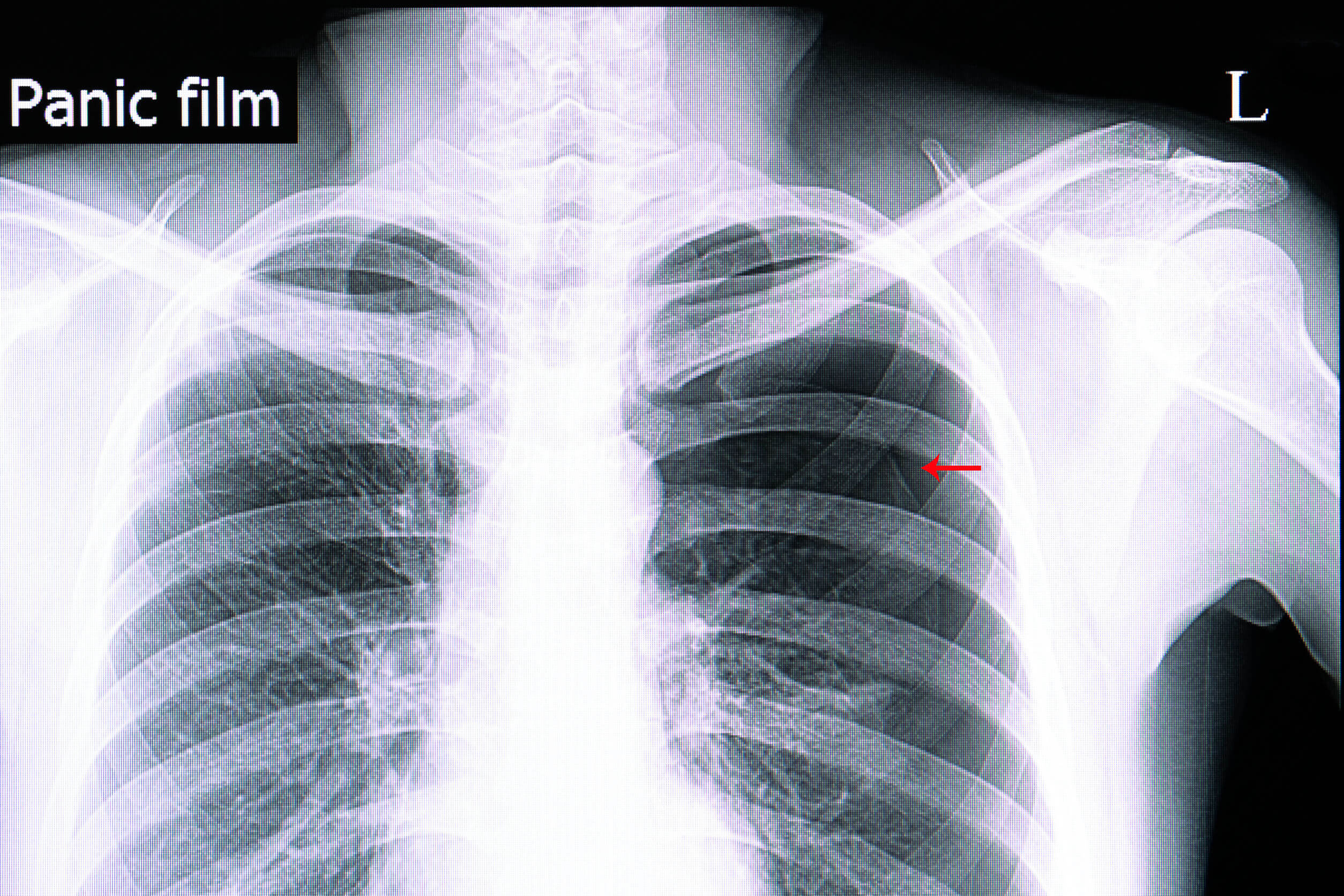
Of these, the plain chest radiograph is the most common. The collapse of both the lung and its vascular structures is usually very evident, despite the fact that in certain patients it could go unnoticed. In the latter case, the use of a CT scan may be beneficial.
Treatment
The goal of the treatment is to remove the air inside the pleural cavity, to promote adequate lung expansion and the restoration of the mechanics of this system. Depending on the case, surgical and non-surgical treatment can be used.
The surgical treatment par excellence is videothoracoscopy, which allows the specialist to locate and close the areas where there’s an air leak. It’s very useful in cases of spontaneous pneumothorax.
In traumatic cases, the insertion of a flexible tube favors the escape of air. However, since recurrence in the form of spontaneous pneumothorax can be high, a surgical procedure is recommended to permanently seal the area of the initial injury.
Pneumothorax, a medical emergency
While some cases of pneumothorax can be managed within a few hours, they can also worsen and cause a life-threatening form called a tension pneumothorax. Therefore, it’ll be necessary to go to the nearest hospital or emergency services if you should notice the aforementioned symptoms.
Pneumothorax is a pathology that causes lung collapse as a consequence of the entry of air into the pleural cavity. It can be caused by several conditions, although traumatic cases are the most frequent in some population groups.
Below, you’ll find a short article on the main characteristics of this condition. Keep reading to find out more!
Anatomy and physiology of the respiratory system
The respiratory system is made up of the airways and the lungs. The former are made up of the larynx, trachea, bronchi, bronchioles, and alveoli. The lungs are made up of highly elastic connective tissue.
Of course, there’s vascular tissue from the heart, and its smaller blood vessels are closely related to the alveoli.
The lungs are located in the chest cavity, and are surrounded by tissue called the pleura. This is divided into two very thin but resistant layers. The one closest to the outer surface of the lungs is called visceral, and the one that’s attached to the chest walls is called parietal.
The space between the two is called the pleural cavity, and it contains a small amount of fluid. Thanks to a complex mechanism related to the pressure difference between the atmosphere and the interior of the lungs, a phenomenon called ventilation occurs.
Thus, human beings are able to inhale oxygen-rich air and exhale air with abundant carbon dioxide, coming from the metabolism of the cells. This process is called gas exchange.
When oxygen reaches the alveoli, it travels to the blood vessels, which drain on the left side of the heart so that blood is distributed to the rest of the body. In the same way, carbon dioxide passes from the small vessels into the alveoli, to enable the body to expel the gas.
Definition and symptoms of pneumothorax
Pneumothorax is a medical emergency caused by the entry of air into the pleural cavity. As we mentioned before, it naturally contains a small amount of fluid. Air can come from both inside and outside the body. In the latter case, the reason is usually chest trauma.
Due to some physical phenomena, the lung on the affected side can collapse. Therefore, medical problems can occur due to a sudden lack of oxygenation. The symptoms are very characteristic, and the most frequent are the following:
- Shortness of breath (dyspnea)
- Increased heart rate (tachycardia)
- Shallow ventilation, but accompanied by increased respiratory rate (tachypnea)
- Dry cough
- Nervousness
- Profuse sweating
Risk factors for pneumothorax
Although a large number of cases of pneumothorax occur as a consequence of open chest trauma, there are some illnesses that increase the probability of suffering primary or spontaneous cases. Some of these conditions are as follows:
- Personal history of pneumothorax, regardless of the cause
- Chronic Obstructive Pulmonary Disease (COPD)
- Chronic smoking
- Need for mechanical ventilation for any pathology. These patients are usually in intensive care units.
According to the World Health Organization (WHO), COPD is a chronic inflammatory disease that affects lung tissue.
It’s more common in patients over 40 years of age, and is usually the consequence of smoking or exposure to biomass smoke. The latter is more frequent in rural communities, where it’s cooked with firewood.
Types and causes
It’s possible to distinguish four types of pneumothorax, the differences of which lie in the pathophysiological mechanisms: traumatic, iatrogenic, related to mechanical ventilation, and spontaneous.
Traumatic
This is usually the most frequent type in young patients, especially those who live in violent areas. Both stab wounds and gunshot wounds are capable of producing a traumatic pneumothorax.
Perforation of the parietal pleura directly results in communication between the exterior and the pleural cavity. Due to the pressure difference, air enters the cavity, causing almost immediate lung collapse.
Iatrogenic and related to mechanical ventilation
The term iatrogenesis is used to designate any medical problem related to a therapeutic act. This can be caused by human error or as an adverse effect of some treatment.
During some procedures such as cannulation of venous access through the subclavian vein, taking a lung biopsy, and thoracentesis, an iatrogenic pneumothorax is possible.
On the other hand, patients with respiratory failure often require mechanical ventilation and hospitalization in intermediate or intensive care units. This makes it possible to artificially provide both the pressure and the oxygen necessary to guarantee adequate lung mechanics.
This can sometimes lead to pulmonary overexpansion and rupture of the alveoli, which can cause the development of pneumothorax and subcutaneous emphysema. This case is also considered an iatrogenic cause.
Spontaneous type
This last type of pneumothorax is the one that tends to cause diagnostic problems, as there’s no obvious cause of the symptoms. It can be subclassified into primary and secondary. The difference lies in the fact that the latter type occurs in patients with previous pulmonary pathologies.
Some of the primary cases occur in patients with previously undiagnosed bullae of the lung. The latter is an air-filled structure that’s usually located in the lung apices. They can form as a result of current or previous infections , as in tuberculosis and some cases of pneumonia.
Diagnosis of pneumothorax
In the vast majority of cases, the diagnosis is clinical. Some complementary studies can be used only in cases where the patient’s life isn’t immediately in danger.
Of these, the plain chest radiograph is the most common. The collapse of both the lung and its vascular structures is usually very evident, despite the fact that in certain patients it could go unnoticed. In the latter case, the use of a CT scan may be beneficial.
Treatment
The goal of the treatment is to remove the air inside the pleural cavity, to promote adequate lung expansion and the restoration of the mechanics of this system. Depending on the case, surgical and non-surgical treatment can be used.
The surgical treatment par excellence is videothoracoscopy, which allows the specialist to locate and close the areas where there’s an air leak. It’s very useful in cases of spontaneous pneumothorax.
In traumatic cases, the insertion of a flexible tube favors the escape of air. However, since recurrence in the form of spontaneous pneumothorax can be high, a surgical procedure is recommended to permanently seal the area of the initial injury.
Pneumothorax, a medical emergency
While some cases of pneumothorax can be managed within a few hours, they can also worsen and cause a life-threatening form called a tension pneumothorax. Therefore, it’ll be necessary to go to the nearest hospital or emergency services if you should notice the aforementioned symptoms.
- González C. Neumotórax espontáneo a tensión. SEMERGEN 2010;36(4):227-229.
- González Fernández AM, Torres Torres AR, Valverde Molina J. Traumatismo torácico, neumotórax, hemoptisis y tromboembolismo pulmonar. Protoc diagn ter pediatr 2017;1:189-209.
- Johnson NN, Toledo A, Endom EE. Pneumothorax, pneumomediastinum and pulmonary embolism. Pediatr Clin N Am. 2010;57:1357-83.
- Porcel J. Neumotórax espontáneo. Medicina Integral 2001;38(1):3-7.
- Roberts DJ, Leigh-Smith S, Faris PD, Ball ChG, Robertson HLBlacmore Ch, et al. Clinical manifestations of tension pneumothorax: protocol for a systemic review and meta-analysis. Syst Rev. 2014;3:3.
- Ossés J, et al. Neumotórax. Revista Medicina Respiratoria 2003;1:35-40.
Este texto se ofrece únicamente con propósitos informativos y no reemplaza la consulta con un profesional. Ante dudas, consulta a tu especialista.


