The Differences Between Veins and Arteries


Escrito y verificado por el biólogo Samuel Antonio Sánchez Amador
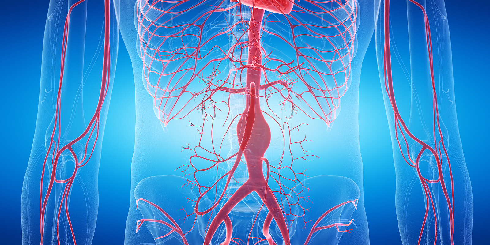
The circulatory system is essential for the conception of life itself. Within an average adult human being, there are about 1.2 gallons of blood (7% of body weight), a liquid connective tissue responsible for transporting oxygen, carbon dioxide, and waste to or from each and every living cell in the body. To understand circulation, it’s necessary to cite the differences between veins and arteries.
Although they seem like structures with the same function, veins and arteries have certain physiological and functional characteristics that make them unique. If you want to know the characteristics of these vital parts of the circulatory system, we encourage you to continue reading.
Overview of the circulatory system
Before exploring the differences between veins and arteries, we find it interesting to cite some key data about the circulatory system and its work in the human body. The informative portal Live Science and other sources help us to bring you the following information of interest:
- The average human being has 1.2 to 1.3 gallons of blood in their body. This is 7-10% of total body weight.
- This system is made up of arteries, arterioles, veins, venules, and capillaries. If all these structures present in the body were put in a row, they would occupy approximately 100,000 kilometers.
- The blood cells par excellence are red blood cells (erythrocytes), and these are responsible for transporting oxygen to all the different tissues in the body. Its proportion in the bloodstream is 1000 times greater than that of leukocytes.
- There are about 5,000,000 red blood cells per cubic millimeter of blood. Each of these cells contains 270,000,000 hemoglobin molecules. For its part, each hemoglobin carries four oxygen molecules.
- The heart is the central organ and is responsible for pumping all the blood through the body. It’s estimated to beat about 2.5 billion times during a life span of 75 years.
In humans, the circulatory system is made up of blood, a set of conduits (veins, arteries, and capillaries, among others), and the driving pump known to all, the heart. It’s crucial to distinguish each structure at a physiological level, as various circulation diseases vary in severity and prognosis according to the affected place.
What are the differences between veins and arteries?
Now that you know what the circulatory system is and some of its essential parts, we’re ready to explore the differences between veins and arteries at various levels: Functional, physiological, anatomical, and pathological. Keep reading.
1. The arteries “go”, while the veins “return”
The National Cancer Institute defines the artery as “a blood vessel that carries blood from the heart to the tissues and organs of the body.” On the other hand, the same institution argues that the vein carries the blood fluid from the organs and tissues to the heart. The first type of conduit goes from the heart outward, while the second goes back to it.
Most arteries carry oxygenated blood, except for the pulmonary and umbilical. On the other hand, most veins carry deoxygenated blood fluid that’s full of carbon dioxide, but there are also exceptions. Although oxygen saturation is the first of the differences between veins and arteries, it’s also the most basic.
Exceptions to the rule
Saying that the arteries carry blood with oxygen (whose interaction with hemoglobin makes it red) and the veins with liquid free of it (blue color) is the most comfortable, but it’s not always true. The following arteries carry deoxygenated blood:
- Pulmonary arteries: The left pulmonary artery and the right pulmonary artery emerge from a single arterial duct on the left side of the heart, and after branching, the resulting capillaries communicate with the venules of the pulmonary system. During this “interaction,” which occurs in the pulmonary alveoli, “gas exchange” occurs.
- Umbilical arteries: During intrauterine development, there are two parallel umbilical arteries, located at the abdominal level. Its job is to transport deoxygenated blood from the fetus to the placenta.
There are also veins that don’t carry deoxygenated blood. These are the following:
- Pulmonary veins: Perform the work that’s opposite to that of the pulmonary arteries, as they collect oxygenated blood from the lungs and carry it to the heart. From there, the oxygen can be distributed to the rest of the tissues.
- Umbilical veins: Umbilical veins carry oxygenated blood from the maternal placenta to the developing fetus. The remnant of these structures after birth (and cutting the umbilical cord) disappears after the first week of life.
The pulmonary veins and arteries follow the opposite premise to the rest of the circulatory system. Therefore, its functionality is treated separately and is analyzed in its own circuit: The pulmonary circulation system. The case of the umbilical circulation is even more special, as its structures degrade and change after the birth of the fetus.
2. The parts of each blood vessel are different
Another difference between veins and arteries lies in their composition at the physiological level. In any case, it should be noted that all blood vessels have a concentric 3-layer arrangement with disparate properties:
- Tunica intima: This is the innermost layer of both arteries and veins. It’s composed of endothelial cells (flattened bodies that line the walls of the vessels), which in turn are in direct contact with the blood flow or lumen of the vessel.
- Tunica media: This layer is made up of smooth muscle cells (cell bodies with lengths between 20 and 500 micrometers and a diameter between 8 and 10 micrometers) and also contains elastin fibers, made up of a protein that provides resistance and elasticity to tissues.
- Tunica adventitia: This is the outer layer of fibrous connective tissue that surrounds all blood vessels. It’s essential for the regulation of the functions of the vascular walls.
Although all blood vessels have the same general structure, it should be noted that each layer can vary in composition and functionality. For example, the tunica media is much more developed in arteries than in veins and is almost imperceptible in blood capillaries.
For this last reason, it is often said that veins are less “muscular” than arteries.
3. Different types of veins and arteries
For now, the differences between arteries and veins are clear: The former usually carry oxygenated blood to the tissues and are more muscular, while the latter bring the blood fluid loaded with CO₂ back to the heart so that this gas can be exchanged with oxygen in the lungs during breathing. Although pulmonary circulation doesn’t follow this rule, it’s applicable to the rest of the processes.
In addition to these baseline distinctions, there are disparities between the two blood vessels in terms of typology. The Healthline medical portal allows us to show you the different types of arteries in the following list:
- Elastic arteries: Elastic arteries are the largest arteries near the heart, such as the aorta or pulmonary. They have a very thick and adaptable middle tunic, as they must stretch when the heart contracts to pump blood.
- Muscular arteries: They differ from elastic arteries by having more smooth muscle and fewer elastic fibers than the aforementioned counterpart. They’re usually medium in size.
- Arterioles: These are the smallest units of this type before reaching the capillary level. For an arteriole to be considered as such, it must have a diameter of less than 40-100 microns. They represent the main points of vascular resistance.
On the other hand, the following types of veins can be recorded:
- Deep veins: As the name suggests, these veins are located far from the most superficial layers of the body. They have one-way valves that prevent blood from backing up, and their muscles help it continue on its way to the heart. They represent a fairly important medical problem, as it’s very difficult to access them in pathological conditions.
- Superficial veins: They’re much closer to the skin and are easily accessible. They have the same type of valves as deep veins, but they aren’t surrounded by muscles. For this reason, these types of venous vessels don’t actively push the blood fluid to the heart and the flow is slower.
- Venules: They’re analogous to arterioles but in the venous area. They’re the first to direct blood to the heart after contact with blood capillaries.
Each blood vessel is divided into three different subtypes, but as you may have seen, the classification system is somewhat different. The types of veins are defined more by their location in terms of “depth,” while the arteries are classified by the composition of their layers (especially the tunica media).
Although this is a standard, there are more qualifying criteria for veins and arteries.
4. Pressure differences

When the heart contracts and pushes blood into the blood vessels, they experience some pressure. One of the most important differences between veins and arteries lies in this parameter, as it can elucidate certain chronic diseases that must be treated.
As indicated by the United States National Library of Medicine, the term blood pressure refers to the force exerted by the blood when it pushes against the walls of the tissues of the arteries when it advances. This concept is divided into 2 values: Systolic (when the heart contracts) and diastolic (when it relaxes).
Systolic blood pressure is higher than diastolic, and the respective normal values are 120/80 millimeters of mercury (mmHg). A person is said to have hypertension when constant pressures hover over or exceed the 140/90 mmHg limit.
On the other hand, venous pressure refers to the force exerted by blood on the walls of the venous vessels as it passes. Its values are much lower than in the arterial variant and, in addition, it’s measured at two different points:
- Central Venous Pressure (PVC): This term refers to the pressure exerted by the blood in the superior vena cava. It reflects the amount of blood that returns to the heart and its average value is between 0 and 5 mmHg.
- Peripheral Venous Pressure (PVP): This value is measured in veins outside the chest (extrathoracic vessels). It’s somewhat higher than the PVC and depends on the location of the analyzed vessel. In general, their values range between 16 mmHg and 5 mmHg.
Blood pressure is always much higher than venous pressure, as the compliance of the veins is much higher (although the blood volume is large). In addition, when we talk about hypertension, we’re always referring to the systolic/diastolic pressure values in the arteries, never in the veins.
More than 40% of the adult population in high-income countries is hypertensive. Up to 37% of patients haven’t received the relevant diagnosis and aren’t aware of their disease.
The differences between veins and arteries: Two sides of the same coin
The differences between veins and arteries are multiple, but can be summed up in a central idea: Arteries carry oxygen-laden blood to the body’s tissues, while veins return CO2-laden blood and metabolic waste to the heart. The pulmonary circulatory system is the exception to this general rule.
Despite their disparities, both types of blood vessels are essential to understanding the functioning of the circulatory system and its possible diseases. Although arteries are best known for one of the most widespread chronic diseases in the world (hypertension), veins are just as important when it comes to understanding blood flow in the body.
The circulatory system is essential for the conception of life itself. Within an average adult human being, there are about 1.2 gallons of blood (7% of body weight), a liquid connective tissue responsible for transporting oxygen, carbon dioxide, and waste to or from each and every living cell in the body. To understand circulation, it’s necessary to cite the differences between veins and arteries.
Although they seem like structures with the same function, veins and arteries have certain physiological and functional characteristics that make them unique. If you want to know the characteristics of these vital parts of the circulatory system, we encourage you to continue reading.
Overview of the circulatory system
Before exploring the differences between veins and arteries, we find it interesting to cite some key data about the circulatory system and its work in the human body. The informative portal Live Science and other sources help us to bring you the following information of interest:
- The average human being has 1.2 to 1.3 gallons of blood in their body. This is 7-10% of total body weight.
- This system is made up of arteries, arterioles, veins, venules, and capillaries. If all these structures present in the body were put in a row, they would occupy approximately 100,000 kilometers.
- The blood cells par excellence are red blood cells (erythrocytes), and these are responsible for transporting oxygen to all the different tissues in the body. Its proportion in the bloodstream is 1000 times greater than that of leukocytes.
- There are about 5,000,000 red blood cells per cubic millimeter of blood. Each of these cells contains 270,000,000 hemoglobin molecules. For its part, each hemoglobin carries four oxygen molecules.
- The heart is the central organ and is responsible for pumping all the blood through the body. It’s estimated to beat about 2.5 billion times during a life span of 75 years.
In humans, the circulatory system is made up of blood, a set of conduits (veins, arteries, and capillaries, among others), and the driving pump known to all, the heart. It’s crucial to distinguish each structure at a physiological level, as various circulation diseases vary in severity and prognosis according to the affected place.
What are the differences between veins and arteries?
Now that you know what the circulatory system is and some of its essential parts, we’re ready to explore the differences between veins and arteries at various levels: Functional, physiological, anatomical, and pathological. Keep reading.
1. The arteries “go”, while the veins “return”
The National Cancer Institute defines the artery as “a blood vessel that carries blood from the heart to the tissues and organs of the body.” On the other hand, the same institution argues that the vein carries the blood fluid from the organs and tissues to the heart. The first type of conduit goes from the heart outward, while the second goes back to it.
Most arteries carry oxygenated blood, except for the pulmonary and umbilical. On the other hand, most veins carry deoxygenated blood fluid that’s full of carbon dioxide, but there are also exceptions. Although oxygen saturation is the first of the differences between veins and arteries, it’s also the most basic.
Exceptions to the rule
Saying that the arteries carry blood with oxygen (whose interaction with hemoglobin makes it red) and the veins with liquid free of it (blue color) is the most comfortable, but it’s not always true. The following arteries carry deoxygenated blood:
- Pulmonary arteries: The left pulmonary artery and the right pulmonary artery emerge from a single arterial duct on the left side of the heart, and after branching, the resulting capillaries communicate with the venules of the pulmonary system. During this “interaction,” which occurs in the pulmonary alveoli, “gas exchange” occurs.
- Umbilical arteries: During intrauterine development, there are two parallel umbilical arteries, located at the abdominal level. Its job is to transport deoxygenated blood from the fetus to the placenta.
There are also veins that don’t carry deoxygenated blood. These are the following:
- Pulmonary veins: Perform the work that’s opposite to that of the pulmonary arteries, as they collect oxygenated blood from the lungs and carry it to the heart. From there, the oxygen can be distributed to the rest of the tissues.
- Umbilical veins: Umbilical veins carry oxygenated blood from the maternal placenta to the developing fetus. The remnant of these structures after birth (and cutting the umbilical cord) disappears after the first week of life.
The pulmonary veins and arteries follow the opposite premise to the rest of the circulatory system. Therefore, its functionality is treated separately and is analyzed in its own circuit: The pulmonary circulation system. The case of the umbilical circulation is even more special, as its structures degrade and change after the birth of the fetus.
2. The parts of each blood vessel are different
Another difference between veins and arteries lies in their composition at the physiological level. In any case, it should be noted that all blood vessels have a concentric 3-layer arrangement with disparate properties:
- Tunica intima: This is the innermost layer of both arteries and veins. It’s composed of endothelial cells (flattened bodies that line the walls of the vessels), which in turn are in direct contact with the blood flow or lumen of the vessel.
- Tunica media: This layer is made up of smooth muscle cells (cell bodies with lengths between 20 and 500 micrometers and a diameter between 8 and 10 micrometers) and also contains elastin fibers, made up of a protein that provides resistance and elasticity to tissues.
- Tunica adventitia: This is the outer layer of fibrous connective tissue that surrounds all blood vessels. It’s essential for the regulation of the functions of the vascular walls.
Although all blood vessels have the same general structure, it should be noted that each layer can vary in composition and functionality. For example, the tunica media is much more developed in arteries than in veins and is almost imperceptible in blood capillaries.
For this last reason, it is often said that veins are less “muscular” than arteries.
3. Different types of veins and arteries
For now, the differences between arteries and veins are clear: The former usually carry oxygenated blood to the tissues and are more muscular, while the latter bring the blood fluid loaded with CO₂ back to the heart so that this gas can be exchanged with oxygen in the lungs during breathing. Although pulmonary circulation doesn’t follow this rule, it’s applicable to the rest of the processes.
In addition to these baseline distinctions, there are disparities between the two blood vessels in terms of typology. The Healthline medical portal allows us to show you the different types of arteries in the following list:
- Elastic arteries: Elastic arteries are the largest arteries near the heart, such as the aorta or pulmonary. They have a very thick and adaptable middle tunic, as they must stretch when the heart contracts to pump blood.
- Muscular arteries: They differ from elastic arteries by having more smooth muscle and fewer elastic fibers than the aforementioned counterpart. They’re usually medium in size.
- Arterioles: These are the smallest units of this type before reaching the capillary level. For an arteriole to be considered as such, it must have a diameter of less than 40-100 microns. They represent the main points of vascular resistance.
On the other hand, the following types of veins can be recorded:
- Deep veins: As the name suggests, these veins are located far from the most superficial layers of the body. They have one-way valves that prevent blood from backing up, and their muscles help it continue on its way to the heart. They represent a fairly important medical problem, as it’s very difficult to access them in pathological conditions.
- Superficial veins: They’re much closer to the skin and are easily accessible. They have the same type of valves as deep veins, but they aren’t surrounded by muscles. For this reason, these types of venous vessels don’t actively push the blood fluid to the heart and the flow is slower.
- Venules: They’re analogous to arterioles but in the venous area. They’re the first to direct blood to the heart after contact with blood capillaries.
Each blood vessel is divided into three different subtypes, but as you may have seen, the classification system is somewhat different. The types of veins are defined more by their location in terms of “depth,” while the arteries are classified by the composition of their layers (especially the tunica media).
Although this is a standard, there are more qualifying criteria for veins and arteries.
4. Pressure differences
When the heart contracts and pushes blood into the blood vessels, they experience some pressure. One of the most important differences between veins and arteries lies in this parameter, as it can elucidate certain chronic diseases that must be treated.
As indicated by the United States National Library of Medicine, the term blood pressure refers to the force exerted by the blood when it pushes against the walls of the tissues of the arteries when it advances. This concept is divided into 2 values: Systolic (when the heart contracts) and diastolic (when it relaxes).
Systolic blood pressure is higher than diastolic, and the respective normal values are 120/80 millimeters of mercury (mmHg). A person is said to have hypertension when constant pressures hover over or exceed the 140/90 mmHg limit.
On the other hand, venous pressure refers to the force exerted by blood on the walls of the venous vessels as it passes. Its values are much lower than in the arterial variant and, in addition, it’s measured at two different points:
- Central Venous Pressure (PVC): This term refers to the pressure exerted by the blood in the superior vena cava. It reflects the amount of blood that returns to the heart and its average value is between 0 and 5 mmHg.
- Peripheral Venous Pressure (PVP): This value is measured in veins outside the chest (extrathoracic vessels). It’s somewhat higher than the PVC and depends on the location of the analyzed vessel. In general, their values range between 16 mmHg and 5 mmHg.
Blood pressure is always much higher than venous pressure, as the compliance of the veins is much higher (although the blood volume is large). In addition, when we talk about hypertension, we’re always referring to the systolic/diastolic pressure values in the arteries, never in the veins.
More than 40% of the adult population in high-income countries is hypertensive. Up to 37% of patients haven’t received the relevant diagnosis and aren’t aware of their disease.
The differences between veins and arteries: Two sides of the same coin
The differences between veins and arteries are multiple, but can be summed up in a central idea: Arteries carry oxygen-laden blood to the body’s tissues, while veins return CO2-laden blood and metabolic waste to the heart. The pulmonary circulatory system is the exception to this general rule.
Despite their disparities, both types of blood vessels are essential to understanding the functioning of the circulatory system and its possible diseases. Although arteries are best known for one of the most widespread chronic diseases in the world (hypertension), veins are just as important when it comes to understanding blood flow in the body.
- 11 surprising facts about the circulatory system, Live Science. Recogido a 18 de septiembre en https://www.livescience.com/39925-circulatory-system-facts-surprising.html
- Definición de arteria, NIH. Recogido a 18 de septiembre en https://www.cancer.gov/espanol/publicaciones/diccionarios/diccionario-cancer/def/arteria
- Artery VS vein, Healthline. Recogido a 18 de septiembre en https://www.healthline.com/health/artery-vs-vein#types-of-veins
- Presión arterial, Medlineplus.gov. Recogido a 18 de septiembre en https://medlineplus.gov/spanish/highbloodpressure.html
Este texto se ofrece únicamente con propósitos informativos y no reemplaza la consulta con un profesional. Ante dudas, consulta a tu especialista.


