Retinal Detachment: Symptoms, Causes and Treatment
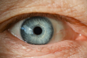
The human eye is made up of various layers, the retina being one of the most important. Vision depends on the ability of the latter to capture the light beams and transmit them through the optic nerve. Are you interested in knowing what are the symptoms, causes and treatment of retinal detachment? Coming up next, we’ll tell you.
The retina is a neurosensory tissue located inside the eyeball. It works through a delicate system of nerve receptors and transducers. We speak of detachment when the retina separates from the posterior wall of the eyeball, altering its function.
Worldwide, 1 positive case is observed for every 10,000 patients per year, which gives an average incidence of 0.01%.
Symptoms of retinal detachment

The clinical manifestations of retinal detachment usually appear quickly and abruptly, unlike other pathologies such as presbyopia. In general, the symptoms depend on the causative agent and the involvement of the retinal layer.
The cardinal symptom is compromised visual acuity, which can range from cloudy vision to vision loss. Similarly, patients with retinal detachment usually have the following symptoms:
- Shadows in the visual field.
- Flashes of light or photopsies.
- Dark floating bodies or myodesopsias.
- Difficulty focusing on objects around you.
In most cases, the person does not experience eye pain. Similarly, light flashes and floating bodies do not always indicate serious visual pathology and are common in elderly patients. However, it is vital to go immediately to the specialist to guarantee timely treatment.
What is the cause?
The pathogenesis involved in retinal detachment is associated with tears or ruptures of the retinal layer, as well as exudative or traction phenomena. They weaken and create gaps between the retina and the wall of the eyeball, which cause detachment.
The most common cause is retraction of the posterior vitreous body. This phenomenon drags part of the retina causing lesions that will induce detachment. Once it is completely separated, it will not work properly, producing blurred vision and dark spots in the visual field.
Similarly, this pathology can be the result of traumatic processes at the skull or eye level. In fact, some studies attribute 20-30% of cases to blunt trauma. In addition, it can also be conditioned by alterations in the formation and development of the eyeball, surgical acts and tumor processes.
Risk factors for retinal detachment
In general, risk factors are associated with phenomena or situations that alter the adhesion and support structure of the retina. They are the result of individual variables, underlying pathologies and conditioning external agents, among which the following have been identified:
- Being over 50 years of age.
- Using lenses for distant objects.
- Glaucoma correction surgery.
- Consumption of medications that reduce the size of the pupil, such as pilocarpine.
- History of tears in the contralateral eye.
- Relatives who have suffered retinal detachment.
- Cataract correction surgery.
- High intensity blows to the head.
- Making great physical effort such as lifting heavy objects.
Similarly, diseases such as preeclampsia, glomerulonephritis, and advanced diabetes increase the risk of developing this condition. In addition, benign and malignant neoplastic disorders in the structures of the eyeball are associated with a higher incidence due to causing retinal weakness.
Diagnosis
The identification of retinal detachment requires a comprehensive assessment by the specialist physician based on the ocular symptoms indicated by the patient during the interrogation. However, the standard test for the diagnosis of this pathology is indirect ophthalmoscopy with previous pupillary dilation.
It allows you to assess all the internal structures of the eye, as well as to determine the integrity and adhesion of the retinal layer. In some patients, vitreous hemorrhages can be seen that make the retina dark. In addition, there is usually the formation of new blood vessels in triggering systemic diseases such as diabetes.
On the other hand, direct ophthalmoscopy can also be of diagnostic utility. However, it can omit retinal injuries or detachments located on the periphery of the eyeball.
Treatment of retinal detachment
Patients diagnosed with this condition should be treated immediately to avoid further damage to the eyeball leading to vision loss.
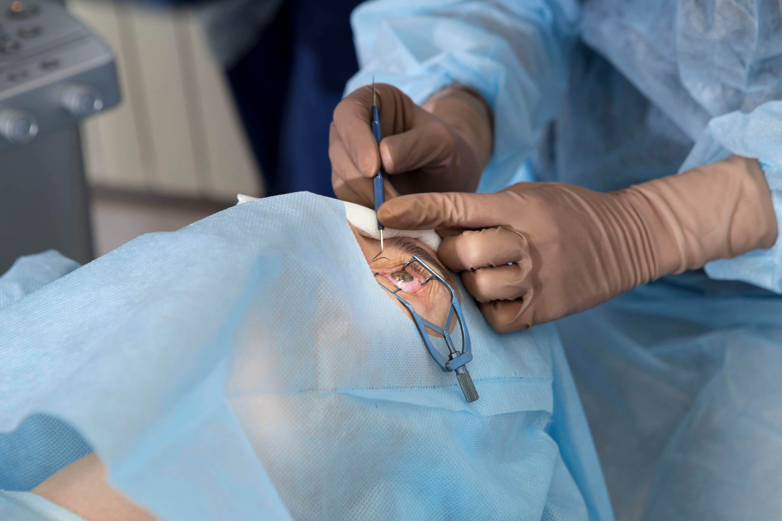
The therapeutic protocol is surgical and aims to solve the structural lesion, with laser or cryotherapy, and place the retina in contact with the posterior surface of the eye.
The most common surgical procedures in the treatment of retinal detachment are the following:
Vitrectomy
The vitreous body is removed and replaced, which retracts the retina by a delicate bubble of oil, gas or air. The latter will be in charge of positioning and holding the retina in place for the time necessary for it to heal properly.
Once the procedure is performed, the patient will not be able to travel to high altitude sites or perform diving activities. They would induce the expansion of the bubble inside the eye, causing a sudden increase in intraocular pressure.
Pneumatic retinopexy
The specialist will place a gas bubble inside the eyeball that will fix the retina to the back wall. After a few days, the eye will produce a liquid that will gradually replace the gas bubble. After the procedure, the doctor will ask to keep the head fixed in a certain position to promote healing.
Scleral loop
A soft rubber band is attached to the outside of the eyeball that will be responsible for pressing inward on the eye. It will allow the detached retina to adhere more easily to the wall. In most cases, this band sits permanently on the eye.
When to go to the doctor?
The prognosis for retinal detachment generally depends on the severity and location of the injury. Surgical resolution usually offers excellent results, however, they depend on the care and timely management of the condition.
Faced with annoying eye symptoms associated with altered visual acuity, you should see a doctor immediately. He will be in charge of providing the necessary help and establishing the care protocol that is required according to the severity of the case.
The human eye is made up of various layers, the retina being one of the most important. Vision depends on the ability of the latter to capture the light beams and transmit them through the optic nerve. Are you interested in knowing what are the symptoms, causes and treatment of retinal detachment? Coming up next, we’ll tell you.
The retina is a neurosensory tissue located inside the eyeball. It works through a delicate system of nerve receptors and transducers. We speak of detachment when the retina separates from the posterior wall of the eyeball, altering its function.
Worldwide, 1 positive case is observed for every 10,000 patients per year, which gives an average incidence of 0.01%.
Symptoms of retinal detachment
The clinical manifestations of retinal detachment usually appear quickly and abruptly, unlike other pathologies such as presbyopia. In general, the symptoms depend on the causative agent and the involvement of the retinal layer.
The cardinal symptom is compromised visual acuity, which can range from cloudy vision to vision loss. Similarly, patients with retinal detachment usually have the following symptoms:
- Shadows in the visual field.
- Flashes of light or photopsies.
- Dark floating bodies or myodesopsias.
- Difficulty focusing on objects around you.
In most cases, the person does not experience eye pain. Similarly, light flashes and floating bodies do not always indicate serious visual pathology and are common in elderly patients. However, it is vital to go immediately to the specialist to guarantee timely treatment.
What is the cause?
The pathogenesis involved in retinal detachment is associated with tears or ruptures of the retinal layer, as well as exudative or traction phenomena. They weaken and create gaps between the retina and the wall of the eyeball, which cause detachment.
The most common cause is retraction of the posterior vitreous body. This phenomenon drags part of the retina causing lesions that will induce detachment. Once it is completely separated, it will not work properly, producing blurred vision and dark spots in the visual field.
Similarly, this pathology can be the result of traumatic processes at the skull or eye level. In fact, some studies attribute 20-30% of cases to blunt trauma. In addition, it can also be conditioned by alterations in the formation and development of the eyeball, surgical acts and tumor processes.
Risk factors for retinal detachment
In general, risk factors are associated with phenomena or situations that alter the adhesion and support structure of the retina. They are the result of individual variables, underlying pathologies and conditioning external agents, among which the following have been identified:
- Being over 50 years of age.
- Using lenses for distant objects.
- Glaucoma correction surgery.
- Consumption of medications that reduce the size of the pupil, such as pilocarpine.
- History of tears in the contralateral eye.
- Relatives who have suffered retinal detachment.
- Cataract correction surgery.
- High intensity blows to the head.
- Making great physical effort such as lifting heavy objects.
Similarly, diseases such as preeclampsia, glomerulonephritis, and advanced diabetes increase the risk of developing this condition. In addition, benign and malignant neoplastic disorders in the structures of the eyeball are associated with a higher incidence due to causing retinal weakness.
Diagnosis
The identification of retinal detachment requires a comprehensive assessment by the specialist physician based on the ocular symptoms indicated by the patient during the interrogation. However, the standard test for the diagnosis of this pathology is indirect ophthalmoscopy with previous pupillary dilation.
It allows you to assess all the internal structures of the eye, as well as to determine the integrity and adhesion of the retinal layer. In some patients, vitreous hemorrhages can be seen that make the retina dark. In addition, there is usually the formation of new blood vessels in triggering systemic diseases such as diabetes.
On the other hand, direct ophthalmoscopy can also be of diagnostic utility. However, it can omit retinal injuries or detachments located on the periphery of the eyeball.
Treatment of retinal detachment
Patients diagnosed with this condition should be treated immediately to avoid further damage to the eyeball leading to vision loss.
The therapeutic protocol is surgical and aims to solve the structural lesion, with laser or cryotherapy, and place the retina in contact with the posterior surface of the eye.
The most common surgical procedures in the treatment of retinal detachment are the following:
Vitrectomy
The vitreous body is removed and replaced, which retracts the retina by a delicate bubble of oil, gas or air. The latter will be in charge of positioning and holding the retina in place for the time necessary for it to heal properly.
Once the procedure is performed, the patient will not be able to travel to high altitude sites or perform diving activities. They would induce the expansion of the bubble inside the eye, causing a sudden increase in intraocular pressure.
Pneumatic retinopexy
The specialist will place a gas bubble inside the eyeball that will fix the retina to the back wall. After a few days, the eye will produce a liquid that will gradually replace the gas bubble. After the procedure, the doctor will ask to keep the head fixed in a certain position to promote healing.
Scleral loop
A soft rubber band is attached to the outside of the eyeball that will be responsible for pressing inward on the eye. It will allow the detached retina to adhere more easily to the wall. In most cases, this band sits permanently on the eye.
When to go to the doctor?
The prognosis for retinal detachment generally depends on the severity and location of the injury. Surgical resolution usually offers excellent results, however, they depend on the care and timely management of the condition.
Faced with annoying eye symptoms associated with altered visual acuity, you should see a doctor immediately. He will be in charge of providing the necessary help and establishing the care protocol that is required according to the severity of the case.
- Jaime Claramunt L. Desprendimiento de retina. Revista Médica Clínica Las Condes. 2010;21(6):956-960.
- Cano Reyes J, Infante Tavio N, González Guerrero L, Fernández Pérez S, Herrera Cutié D. Desprendimiento de retina: una revisión bibliográfica necesaria. MEDISAN. 2015;19( 1 ): 78-87.
- Molina Cisneros C, Alemañy Rubio E, Triana Casado I, González Rodríguez L. Evaluación y conducta recomendada en presencia de precursores vítreorretinianos del desprendimiento de retina. Rev Cubana Oftalmol. 2013; 26( 3 ): 482-499.
- Steel D. Retinal detachment. BMJ Clin Evid. 2014 Mar 3;2014:0710.
- Amer R, Nalcı H, Yalçındağ N. Exudative retinal detachment. Surv Ophthalmol. 2017 Nov-Dec;62(6):723-769.
- Hoogewoud F, Chronopoulos A, Varga Z, Souteyrand G, Thumann G, Schutz JS. Traumatic retinal detachment–the difficulty and importance of correct diagnosis. Surv Ophthalmol. 2016 Mar-Apr;61(2):156-63.
Este texto se ofrece únicamente con propósitos informativos y no reemplaza la consulta con un profesional. Ante dudas, consulta a tu especialista.


