Blood Clots: Whar Are the Warning Signs?
Blood clots can be very harmful if not treated correctly. For this reason, it’s very important to know how to detect direct or indirect signs of thrombosis, which in most cases can be easily detected.
We have prepared the following article so that you can learn to detect four basic signs of any case of clot formation, taking deep vein thrombosis as a representative example. Read on to find out!
Why do blood clots form?
Blood is in a liquid state due to a delicate balance between two different systems. First, those that promote coagulation (procoagulants) and second, those that prevent it (anticoagulants).
This is important in many situations of daily life, such as falls. Can you imagine a small blow to the knee making you bleed uncontrollably? Something similar is suffered by patients with untreated hemophilia, a bleeding disorder.
In humans and mammals in general, blood clots are made of platelets and fibrin. The latter is a substance that is produced and accumulated thanks to the action of proteins called coagulation factors.
Why are blood clots so dangerous?
The body is full of vessels that are responsible for carrying oxygenated blood to the tissues and removing waste. With the exception of a few cells, the blood supply goes everywhere. If a sizable clot becomes lodged near any major organ, the supply of oxygen and nutrients would be cut off.
This, in the long run, could lead to the irreversible loss of the cells and even death. By way of an example, if blood clots settle in the middle cerebral artery, the direct consequence would be the development of a cerebrovascular disease, known as a cerebrovascular accident or ACV, with serious consequences.

What diseases can form blood clots?
Many disorders can cause this type of problem. Some examples are nephrotic syndrome, systemic lupus erythematosus, atrial fibrillation, and diseases involving coagulation factors.
There’s a medical condition known as deep vein thrombosis (DVT). Due to its easy identification that attracts the attention of any patient, it’s useful to use it as a model of the signs that can warn us of the presence of blood clots. It affects the elderly a lot and its triggers include obesity, old age, physical disability, and cancer.
Warning signs: when you should go to the doctor
Below you will find a list of the main elements that you must take into account in order to detect the possible presence of blood clots in your legs:
- Local pain
- Volume increase (swelling)
- Increase in temperature
- Changes in skin coloration
It’s important to note that most of these signs usually occur together. Just one of the symptoms may indicate that the process is just beginning.
1. Local pain
As we mentioned, obstructing blood flow to a certain tissue decreases the number of nutrients that reach it. Of course, waste substances will begin to accumulate and, in many cases, these are toxic.
Because the free nerve endings responsible for causing pain are sensitive to chemical compounds, these substances cause an uncomfortable sensation in the area of the ischemia (a term that refers to the deficit of blood flow in a certain tissue).
One of the operations that doctors perform is to assess whether, when lying on your back, the dorsiflexion of the foot causes calf pain. This is known as the Homans sign, although its evaluation is somewhat outdated thanks to the development of ultrasonography.
2. Increase in volume (swelling)
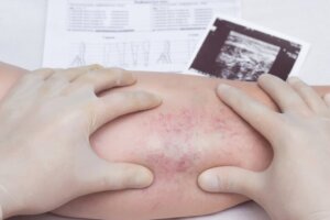
Perhaps this is the most important distinctive element to take into account. When the thrombosis is superficial, you may find a very well-defined, hard, and cold mass that corresponds to the clot. But, when it’s deep there’s an increase in volume.
In addition, blood clots exert pressure within the vessel, causing an obstructive process that prevents proper circulation. This leads to fluid being distributed or leaking into the interstitium, the space between cells in different tissues and the lumen of blood vessels.
3. Increase in temperature
This is a sure sign that an inflammatory phenomenon is taking place within the tissue. Toxic substances that are released into the organs by the abrupt interruption of blood flow also have pro-inflammatory properties. Beyond causing an immediate serious consequence, it can serve as a warning sign.
The easiest way to detect it is to touch the affected area with the same hand and compare it with the other leg. You will notice the temperature difference immediately.
4. Changes in coloration
The same inflammatory phenomenon can cause increased blood flow in adjacent vessels. This makes a lot of sense; it’s a way for the body to guarantee a supply of nutrients when problems occur.
It would be the perfect solution if it weren’t for the fact that these vessels don’t usually directly reach the organ involved, especially when it’s very large and it’s difficult to replace its functions in a short time. Usually, the color you’ll find is red, which is compatible with all of the changes just mentioned.
Which doctor to go to in cases of blood clots?
If you have any of the aforementioned symptoms, there are a wide variety of medical specialties that can offer you care. In principle, the family or emergency doctor can easily detect what is happening and institute immediate treatment. If necessary, you may be referred to another specialist for continued care.
The other specialists who are capable of diagnosing and treating a case of DVT are cardiologists, hematologists, and cardiovascular surgeons. Of course, the management within each hospital center is very particular and, in most cases, may require interventions or multidisciplinary approaches. The most important thing is to never stay at home!

What complementary studies can the doctor request?
The initial clinical evaluation can be complemented with imaging and laboratory studies. This makes it possible to complete the diagnosis and determine how advanced the disease is. The results usually guide the approach.
It’s common to request a complete blood analysis (which includes hematology, lipid profile and coagulation factors) and at least one Doppler ultrasound, in order to identify the position, extension and risks associated with the clot.
What should take into account when I suspect blood clots?
If you have an episode of deep vein thrombosis, it’s recommended that you see a doctor immediately. Your condition will be evaluated, including a possible hospitalization and administration of medications, such as enoxaparin.
It’s very likely that you’ll need to use compression stockings that encourage venous circulation, in addition to contributing to the reduction of inflammation, local edema, and pain. Of course, depending on each case, oral anticoagulant treatment may be prescribed. This is done with the aim of preventing new episodes in the future.
Blood clots can be very harmful if not treated correctly. For this reason, it’s very important to know how to detect direct or indirect signs of thrombosis, which in most cases can be easily detected.
We have prepared the following article so that you can learn to detect four basic signs of any case of clot formation, taking deep vein thrombosis as a representative example. Read on to find out!
Why do blood clots form?
Blood is in a liquid state due to a delicate balance between two different systems. First, those that promote coagulation (procoagulants) and second, those that prevent it (anticoagulants).
This is important in many situations of daily life, such as falls. Can you imagine a small blow to the knee making you bleed uncontrollably? Something similar is suffered by patients with untreated hemophilia, a bleeding disorder.
In humans and mammals in general, blood clots are made of platelets and fibrin. The latter is a substance that is produced and accumulated thanks to the action of proteins called coagulation factors.
Why are blood clots so dangerous?
The body is full of vessels that are responsible for carrying oxygenated blood to the tissues and removing waste. With the exception of a few cells, the blood supply goes everywhere. If a sizable clot becomes lodged near any major organ, the supply of oxygen and nutrients would be cut off.
This, in the long run, could lead to the irreversible loss of the cells and even death. By way of an example, if blood clots settle in the middle cerebral artery, the direct consequence would be the development of a cerebrovascular disease, known as a cerebrovascular accident or ACV, with serious consequences.
What diseases can form blood clots?
Many disorders can cause this type of problem. Some examples are nephrotic syndrome, systemic lupus erythematosus, atrial fibrillation, and diseases involving coagulation factors.
There’s a medical condition known as deep vein thrombosis (DVT). Due to its easy identification that attracts the attention of any patient, it’s useful to use it as a model of the signs that can warn us of the presence of blood clots. It affects the elderly a lot and its triggers include obesity, old age, physical disability, and cancer.
Warning signs: when you should go to the doctor
Below you will find a list of the main elements that you must take into account in order to detect the possible presence of blood clots in your legs:
- Local pain
- Volume increase (swelling)
- Increase in temperature
- Changes in skin coloration
It’s important to note that most of these signs usually occur together. Just one of the symptoms may indicate that the process is just beginning.
1. Local pain
As we mentioned, obstructing blood flow to a certain tissue decreases the number of nutrients that reach it. Of course, waste substances will begin to accumulate and, in many cases, these are toxic.
Because the free nerve endings responsible for causing pain are sensitive to chemical compounds, these substances cause an uncomfortable sensation in the area of the ischemia (a term that refers to the deficit of blood flow in a certain tissue).
One of the operations that doctors perform is to assess whether, when lying on your back, the dorsiflexion of the foot causes calf pain. This is known as the Homans sign, although its evaluation is somewhat outdated thanks to the development of ultrasonography.
2. Increase in volume (swelling)
Perhaps this is the most important distinctive element to take into account. When the thrombosis is superficial, you may find a very well-defined, hard, and cold mass that corresponds to the clot. But, when it’s deep there’s an increase in volume.
In addition, blood clots exert pressure within the vessel, causing an obstructive process that prevents proper circulation. This leads to fluid being distributed or leaking into the interstitium, the space between cells in different tissues and the lumen of blood vessels.
3. Increase in temperature
This is a sure sign that an inflammatory phenomenon is taking place within the tissue. Toxic substances that are released into the organs by the abrupt interruption of blood flow also have pro-inflammatory properties. Beyond causing an immediate serious consequence, it can serve as a warning sign.
The easiest way to detect it is to touch the affected area with the same hand and compare it with the other leg. You will notice the temperature difference immediately.
4. Changes in coloration
The same inflammatory phenomenon can cause increased blood flow in adjacent vessels. This makes a lot of sense; it’s a way for the body to guarantee a supply of nutrients when problems occur.
It would be the perfect solution if it weren’t for the fact that these vessels don’t usually directly reach the organ involved, especially when it’s very large and it’s difficult to replace its functions in a short time. Usually, the color you’ll find is red, which is compatible with all of the changes just mentioned.
Which doctor to go to in cases of blood clots?
If you have any of the aforementioned symptoms, there are a wide variety of medical specialties that can offer you care. In principle, the family or emergency doctor can easily detect what is happening and institute immediate treatment. If necessary, you may be referred to another specialist for continued care.
The other specialists who are capable of diagnosing and treating a case of DVT are cardiologists, hematologists, and cardiovascular surgeons. Of course, the management within each hospital center is very particular and, in most cases, may require interventions or multidisciplinary approaches. The most important thing is to never stay at home!
What complementary studies can the doctor request?
The initial clinical evaluation can be complemented with imaging and laboratory studies. This makes it possible to complete the diagnosis and determine how advanced the disease is. The results usually guide the approach.
It’s common to request a complete blood analysis (which includes hematology, lipid profile and coagulation factors) and at least one Doppler ultrasound, in order to identify the position, extension and risks associated with the clot.
What should take into account when I suspect blood clots?
If you have an episode of deep vein thrombosis, it’s recommended that you see a doctor immediately. Your condition will be evaluated, including a possible hospitalization and administration of medications, such as enoxaparin.
It’s very likely that you’ll need to use compression stockings that encourage venous circulation, in addition to contributing to the reduction of inflammation, local edema, and pain. Of course, depending on each case, oral anticoagulant treatment may be prescribed. This is done with the aim of preventing new episodes in the future.
- Flores-Rivera, Ramírez-Morales, Meza-Márquez, Nava-López. Fisiología de la coagulación. Revista Mexicana de Anestesiología 2014;(37)2:S382-S386.
- Santos, AJ Trujillo. “Tratamiento de la trombosis venosa profunda de extremidades inferiores.” Revista Clínica Española (2020).
- Botella G, Gómez L. Nuevos criterios para el diagnóstico y tratamiento de la trombosis venosa profunda de los miembros inferiores. Anales de Medicina Interna 2004;(21)8:400-407
- Rosas J, Ríos M. Evolución ecográfica de la trombosis venosa profunda en pacientes con trombólisis farmacológica. Anales de Radiología México 2010;(2):76-79.
- Murillo, Carlos Martínez. “Mecanismos de activación de la coagulación.” Revista Médica del Instituto Mexicano del Seguro Social 44.S2 (2006): 51-58.
- Enríquez-Vega, Elizabeth, José Halabe-Cherem, and Janet Tanus-Hajjc. “Diagnóstico de la trombosis venosa profunda.” Gaceta Médica de México 143.S1 (2007): 15-17.
- Camm, A. John, et al. “Guías de práctica clínica para el manejo de la fibrilación auricular.” Revista española de Cardiología 63.12 (2010): 1483.
Este texto se ofrece únicamente con propósitos informativos y no reemplaza la consulta con un profesional. Ante dudas, consulta a tu especialista.


