The 3 Skin Layers: Characteristics and Functions

Escrito y verificado por el biólogo Samuel Antonio Sánchez Amador
The skin is the largest organ in the human body, since in an average adult it has an approximate surface area of 2 square meters (6.5 sq feet) and an estimated weight of 5 kilos (11 pounds). It’s divided into 3 layers and its main function is to protect us against injuries and pathogenic microorganisms, but it also has other vital roles in order to ensure our survival.
The skin is quite simply the protective boundary that protects the body from the external environment. However, the nature of this organ varies considerably according to the area we’re talking about. The skin on the soles of the feet is totally different from the skin on the eyelids, for example. If you’d like to get to know everything about the amazing world of skin, read on!
The skin: the first biological barrier
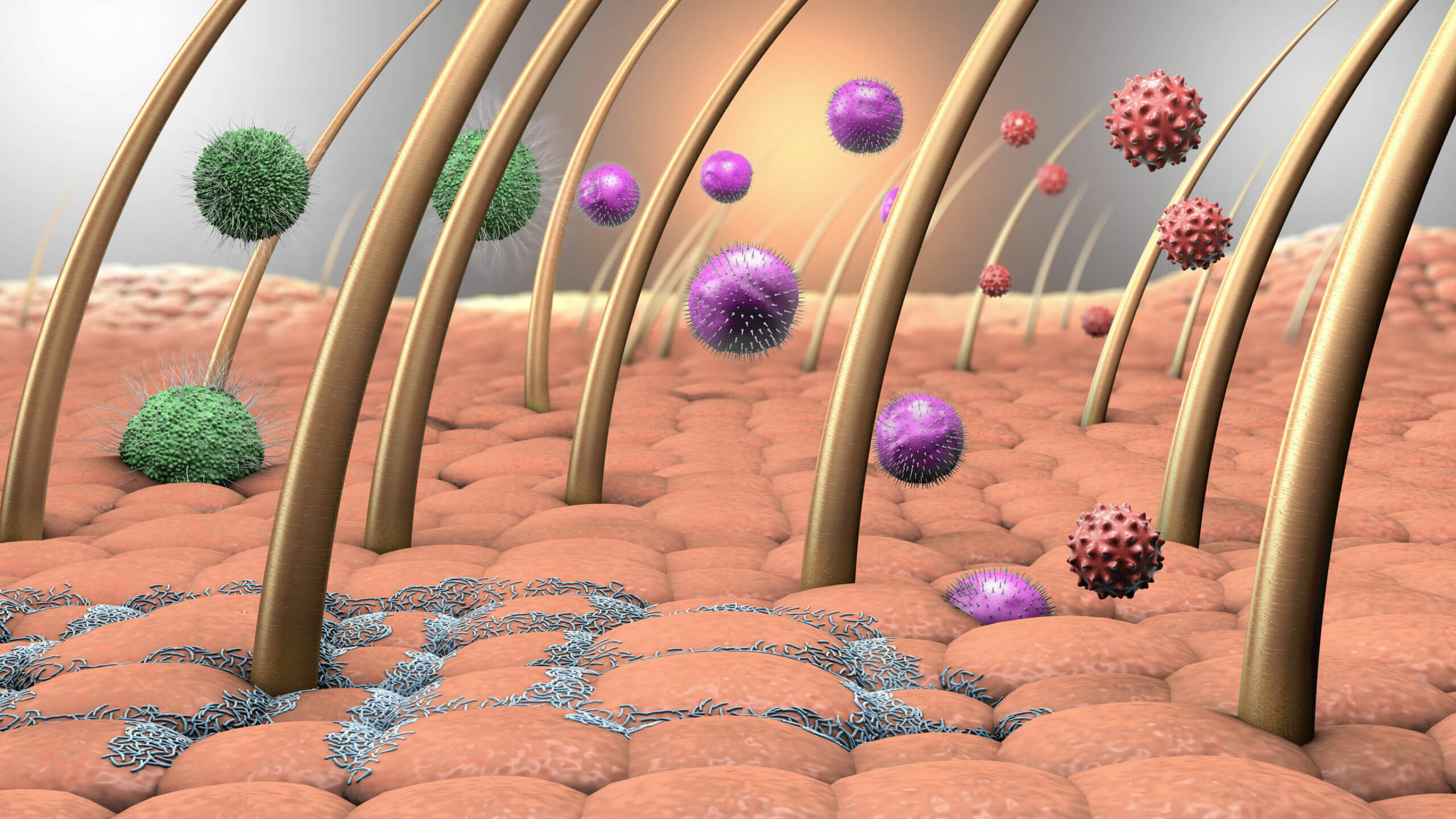
For several decades, we have seen that human beings have two types of immune system: the innate and the acquired. The first is the one we were born with, which protects us in a non-specific way from possible external pathogens and intrinsic processes, such as the appearance of cancer cells or the entry of allergens into the respiratory system.
In general, macrophages and neutrophils are associated with the function of innate immunity. These are types of white blood cells that surround and capture pathogens based on their antigens. However, few people know that human skin is the first physical, chemical, and biological barrier to develop.
First of all, human skin is dry, due to the presence of keratin. This makes bacterial proliferation impossible, and so it’s common for infections to settle in the mucous membranes. It also has a slightly acidic pH due to the presence of sweat, which prevents the proliferation of many microorganisms.
The specialized cell types travel quickly to the skin when it’s hurt, in order to prevent pathogens from infiltrating through wounds and friction, as indicated by the Immunomedia portal. In short: the immune capacity of the skin is invaluable, both physically, chemically and biologically.
Many bacteria grow on the surface of the skin, preventing harmful microorganisms from settling on it.
Other functions of the skin
Despite its importance for the immune system, the skin has many more benefits. The MSD Manuals portal and other sources help us to summarize the most relevant ones in the list:
- Protection of the body against trauma.
- Corporal temperature regulation.
- Maintenance of electrolyte balance.
- Stimulus uptake: here are the nociceptors, nerve cell endings that send the stimulus signal to essential areas of the central nervous system.
- Active intervention in the synthesis of vitamin D: various studies show that the sun’s rays stimulate the transformation of 7-dehydrocholesterol in the epidermis to precolecalciferol, which is converted to vitamin D3 by a heat-mediated reaction.
These are some of the most important features of the skin, but by no means are they the only ones. Thanks to our skin, we remain in a homeostatic balance, that is, stable with respect to changes in the environment. To a large extent, our individuality as a species is highlighted by this organ.
The 3 layers of the skin and their importance
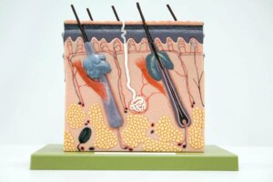
The basic histological structure that we’re going to show you next is common to all vertebrates, including humans. This is divided into 3 general layers: the epidermis, the dermis, and the subcutaneous tissue, also known as the hypodermis.
1. Epidermis: the outermost layer of the skin
The epidermis is the flat, multi-layered epithelium (with more than one layer of cells) that covers the entire body surface. The thickness of this tissue varies depending on the area evaluated. On the eyelid, it doesn’t exceed 0.1 mm, while on the palms and soles it reaches up to 1.5 mm in thickness.
The main cell in this tissue is the keratinocyte, constituting 95% of the cell bodies included here. As indicated by the Eucerin portal, several layers can be distinguished within the epidermis. These are as follows, from bottom to top:
- Basal layer: The innermost layer of all, where keratinocytes are produced.
- Spinous stratum: Here the keratinocytes produce keratin, adopting a spindle shape, hence the name spinous.
- Granular layer: This is where the keratinization begins.
- Lucid layer: This is present in the thickest areas of the skin (such as plants), but not in thin areas. It consists of 3 to 5 rows of flat, clear, dead cells that still contain enzymatic activity.
- Horny layer: The outermost layer of the epidermis, with which we come into contact with the environment and all the inclemency that this entails. This layer of tissue is made up of 15-20 cell layers, and the last one suffers desquamation. The whole strata is replaced every 30 days.
Beyond keratinocytes
Although keratinocytes (living or dead) dominate the epidermis, there are other cell bodies too. One of them is the melanocyte, which is responsible for producing melanin. This compound is essential, as it protects us against the damaging effects of the sun’s rays.
Other structures are Langerhans cells and Merkel cells. The former act as macrophages, since they surround pathogens in a generic way. Merkel cells, for their part, play an essential role as receptors for touch and are found in the basal layers of the epidermis.
Every minute of the day we lose 30,000 to 40,000 skin cells. The regeneration speed of this tissue is dizzying.
2. Dermis
According to the Infermera Virtual portal, the dermis is made up of connective tissue, containing type I collagen fibers and elastic fibers. It’s located below the epidermis and is in contact with it thanks to the dermo-epidermal junction, a 100 nm thick structure that joins both fronts.
The cells of the dermis are much more difficult to approach, as we’re facing a much more heterogeneous mass: fibroblasts, macrophages, mast cells, adipocytes, nerves, blood vessels, glands, and many other structures.
Like the epidermis, the dermis is made up of more than one layer. In this case, however, we will only describe 2 layers:
- Papillary layer: The outermost layer, in contact with the epidermis. It’s a superficial zone of loose connective tissue that contacts the basement membrane. It’s formed by a series of elevations or ridges known as dermal papillae, rich in the aforementioned cellular elements.
- Reticular layer: The widest layer of the dermis, with a variable thickness depending on the section of the skin consulted. It’s a dense irregular connective tissue, rich in type I collagen fibers.
Of all the cell types mentioned, the fibroblast draws our attention. According to the National Cancer Institute of the United States of America, the fibroblast can be defined as a connective tissue cell that synthesizes collagen, a supportive fiber-forming protein that allows several structures to be held together.
Other cell types of the dermis
Some portals already mentioned above show us the peculiarities of other structures within the dermis. In summary, we’re going to tell you about some of them:
- Nerve endings: These detect the stimuli of pain, touch, pressure, and outside temperature. In patients who have damaged nerve endings, hyperalgesia and allodynia may occur. In the first, the painful stimuli become unbearable, and, in the second, things hurt that wouldn’t normally cause pain under normal conditions.
- Sweat glands: These produce sweat in response to temperature and stress. This liquid is made up of water, sodium, and other chemicals. Its main function is thermoregulation and the elimination of toxins.
- Sebaceous glands: These produce sebum in the hair follicles. When the outlet of these glands becomes blocked, those annoying blackheads, known as comedones, appear.
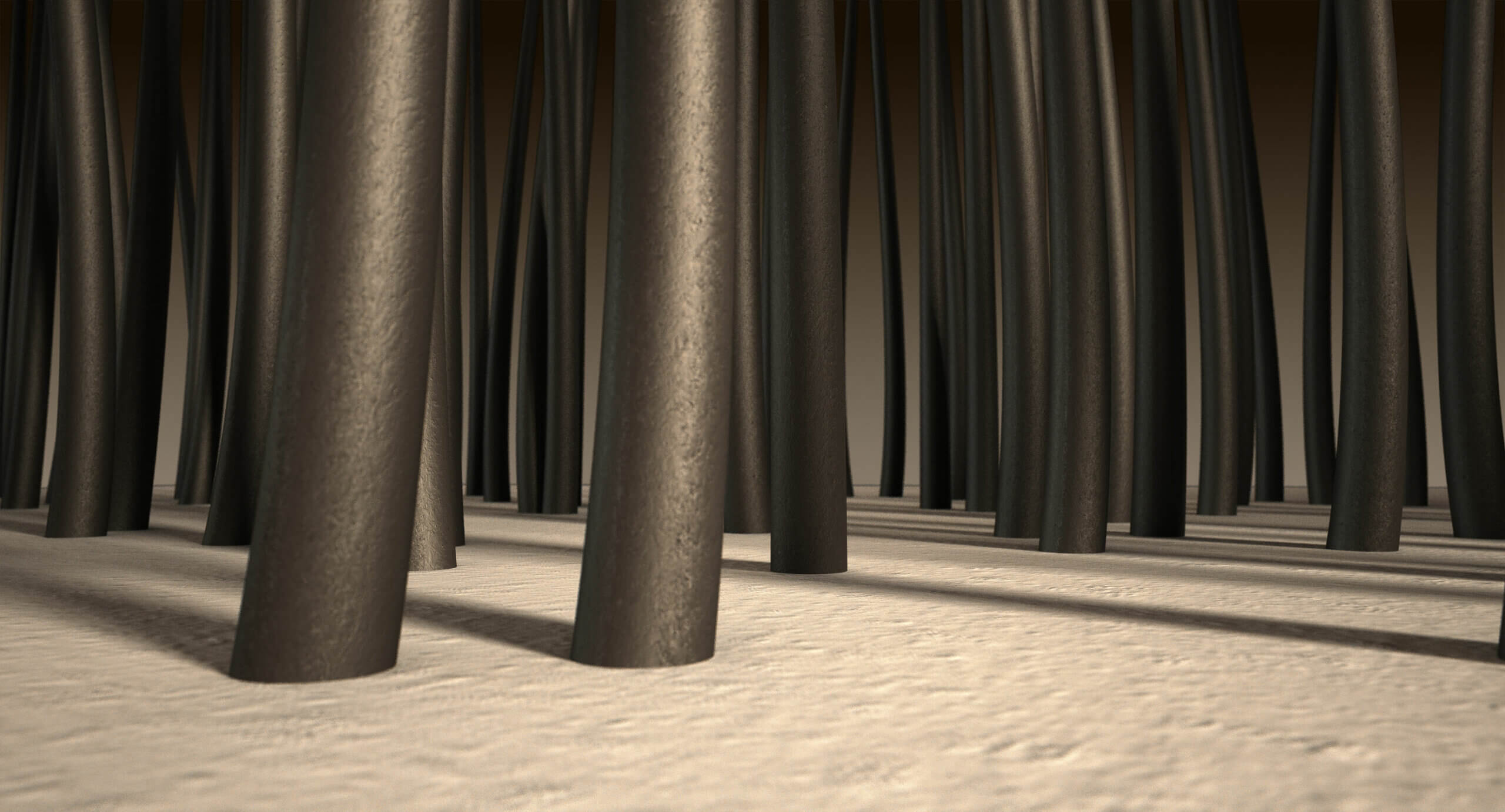
- Hair follicles: These produce the different types of hair that cover the body. A dark-haired person has, on the head alone, an average of about 100,000 hairs.
- Blood vessels: These nourish each and every one of the aforementioned cellular components. They also help thermoregulation, since their expansion or contraction promotes the loss or preservation of heat in the body.
3. Hypodermis
As indicated by the Hospital Alemán medical portal, the hypodermis is the subcutaneous tissue that is used to store fat. For this reason, it’s also known as fatty subcutaneous tissue. The hypodermis is located below the dermal reticular layer and the dominant cell type is the adipocyte.
Adipocytes are cells that are specialized in the production and storage of fats. In addition, they form a fabric that acts as a “mattress”, as it’s very flexible. This protects us from shocks and other types of stress that we may suffer.
When adipocytes increase disproportionately in size due to excess lipid storage, weight gain occurs. On the other hand, during a diet, they decrease in volume, since they release compounds to nourish the cells at a metabolic level.
White adipocytes represent 15-20% of the total fat of an adult, a not inconsiderable percentage.
Summary
We’ve given a brief summary about the three main layers of the skin, all of them subdivided into their corresponding layers, but there’s still enough left to write an entire book! As we’ve said, the skin is one of the largest and most important organs in the body.
This incredible and complex structure is responsible for protecting us against external stress and environmental harshness, and allowing thermoregulation. It’s also responsible for excreting toxins and a host of other functionalities. Without this thin cell layer, life as we know it today would be impossible.
The skin is the largest organ in the human body, since in an average adult it has an approximate surface area of 2 square meters (6.5 sq feet) and an estimated weight of 5 kilos (11 pounds). It’s divided into 3 layers and its main function is to protect us against injuries and pathogenic microorganisms, but it also has other vital roles in order to ensure our survival.
The skin is quite simply the protective boundary that protects the body from the external environment. However, the nature of this organ varies considerably according to the area we’re talking about. The skin on the soles of the feet is totally different from the skin on the eyelids, for example. If you’d like to get to know everything about the amazing world of skin, read on!
The skin: the first biological barrier
For several decades, we have seen that human beings have two types of immune system: the innate and the acquired. The first is the one we were born with, which protects us in a non-specific way from possible external pathogens and intrinsic processes, such as the appearance of cancer cells or the entry of allergens into the respiratory system.
In general, macrophages and neutrophils are associated with the function of innate immunity. These are types of white blood cells that surround and capture pathogens based on their antigens. However, few people know that human skin is the first physical, chemical, and biological barrier to develop.
First of all, human skin is dry, due to the presence of keratin. This makes bacterial proliferation impossible, and so it’s common for infections to settle in the mucous membranes. It also has a slightly acidic pH due to the presence of sweat, which prevents the proliferation of many microorganisms.
The specialized cell types travel quickly to the skin when it’s hurt, in order to prevent pathogens from infiltrating through wounds and friction, as indicated by the Immunomedia portal. In short: the immune capacity of the skin is invaluable, both physically, chemically and biologically.
Many bacteria grow on the surface of the skin, preventing harmful microorganisms from settling on it.
Other functions of the skin
Despite its importance for the immune system, the skin has many more benefits. The MSD Manuals portal and other sources help us to summarize the most relevant ones in the list:
- Protection of the body against trauma.
- Corporal temperature regulation.
- Maintenance of electrolyte balance.
- Stimulus uptake: here are the nociceptors, nerve cell endings that send the stimulus signal to essential areas of the central nervous system.
- Active intervention in the synthesis of vitamin D: various studies show that the sun’s rays stimulate the transformation of 7-dehydrocholesterol in the epidermis to precolecalciferol, which is converted to vitamin D3 by a heat-mediated reaction.
These are some of the most important features of the skin, but by no means are they the only ones. Thanks to our skin, we remain in a homeostatic balance, that is, stable with respect to changes in the environment. To a large extent, our individuality as a species is highlighted by this organ.
The 3 layers of the skin and their importance
The basic histological structure that we’re going to show you next is common to all vertebrates, including humans. This is divided into 3 general layers: the epidermis, the dermis, and the subcutaneous tissue, also known as the hypodermis.
1. Epidermis: the outermost layer of the skin
The epidermis is the flat, multi-layered epithelium (with more than one layer of cells) that covers the entire body surface. The thickness of this tissue varies depending on the area evaluated. On the eyelid, it doesn’t exceed 0.1 mm, while on the palms and soles it reaches up to 1.5 mm in thickness.
The main cell in this tissue is the keratinocyte, constituting 95% of the cell bodies included here. As indicated by the Eucerin portal, several layers can be distinguished within the epidermis. These are as follows, from bottom to top:
- Basal layer: The innermost layer of all, where keratinocytes are produced.
- Spinous stratum: Here the keratinocytes produce keratin, adopting a spindle shape, hence the name spinous.
- Granular layer: This is where the keratinization begins.
- Lucid layer: This is present in the thickest areas of the skin (such as plants), but not in thin areas. It consists of 3 to 5 rows of flat, clear, dead cells that still contain enzymatic activity.
- Horny layer: The outermost layer of the epidermis, with which we come into contact with the environment and all the inclemency that this entails. This layer of tissue is made up of 15-20 cell layers, and the last one suffers desquamation. The whole strata is replaced every 30 days.
Beyond keratinocytes
Although keratinocytes (living or dead) dominate the epidermis, there are other cell bodies too. One of them is the melanocyte, which is responsible for producing melanin. This compound is essential, as it protects us against the damaging effects of the sun’s rays.
Other structures are Langerhans cells and Merkel cells. The former act as macrophages, since they surround pathogens in a generic way. Merkel cells, for their part, play an essential role as receptors for touch and are found in the basal layers of the epidermis.
Every minute of the day we lose 30,000 to 40,000 skin cells. The regeneration speed of this tissue is dizzying.
2. Dermis
According to the Infermera Virtual portal, the dermis is made up of connective tissue, containing type I collagen fibers and elastic fibers. It’s located below the epidermis and is in contact with it thanks to the dermo-epidermal junction, a 100 nm thick structure that joins both fronts.
The cells of the dermis are much more difficult to approach, as we’re facing a much more heterogeneous mass: fibroblasts, macrophages, mast cells, adipocytes, nerves, blood vessels, glands, and many other structures.
Like the epidermis, the dermis is made up of more than one layer. In this case, however, we will only describe 2 layers:
- Papillary layer: The outermost layer, in contact with the epidermis. It’s a superficial zone of loose connective tissue that contacts the basement membrane. It’s formed by a series of elevations or ridges known as dermal papillae, rich in the aforementioned cellular elements.
- Reticular layer: The widest layer of the dermis, with a variable thickness depending on the section of the skin consulted. It’s a dense irregular connective tissue, rich in type I collagen fibers.
Of all the cell types mentioned, the fibroblast draws our attention. According to the National Cancer Institute of the United States of America, the fibroblast can be defined as a connective tissue cell that synthesizes collagen, a supportive fiber-forming protein that allows several structures to be held together.
Other cell types of the dermis
Some portals already mentioned above show us the peculiarities of other structures within the dermis. In summary, we’re going to tell you about some of them:
- Nerve endings: These detect the stimuli of pain, touch, pressure, and outside temperature. In patients who have damaged nerve endings, hyperalgesia and allodynia may occur. In the first, the painful stimuli become unbearable, and, in the second, things hurt that wouldn’t normally cause pain under normal conditions.
- Sweat glands: These produce sweat in response to temperature and stress. This liquid is made up of water, sodium, and other chemicals. Its main function is thermoregulation and the elimination of toxins.
- Sebaceous glands: These produce sebum in the hair follicles. When the outlet of these glands becomes blocked, those annoying blackheads, known as comedones, appear.
- Hair follicles: These produce the different types of hair that cover the body. A dark-haired person has, on the head alone, an average of about 100,000 hairs.
- Blood vessels: These nourish each and every one of the aforementioned cellular components. They also help thermoregulation, since their expansion or contraction promotes the loss or preservation of heat in the body.
3. Hypodermis
As indicated by the Hospital Alemán medical portal, the hypodermis is the subcutaneous tissue that is used to store fat. For this reason, it’s also known as fatty subcutaneous tissue. The hypodermis is located below the dermal reticular layer and the dominant cell type is the adipocyte.
Adipocytes are cells that are specialized in the production and storage of fats. In addition, they form a fabric that acts as a “mattress”, as it’s very flexible. This protects us from shocks and other types of stress that we may suffer.
When adipocytes increase disproportionately in size due to excess lipid storage, weight gain occurs. On the other hand, during a diet, they decrease in volume, since they release compounds to nourish the cells at a metabolic level.
White adipocytes represent 15-20% of the total fat of an adult, a not inconsiderable percentage.
Summary
We’ve given a brief summary about the three main layers of the skin, all of them subdivided into their corresponding layers, but there’s still enough left to write an entire book! As we’ve said, the skin is one of the largest and most important organs in the body.
This incredible and complex structure is responsible for protecting us against external stress and environmental harshness, and allowing thermoregulation. It’s also responsible for excreting toxins and a host of other functionalities. Without this thin cell layer, life as we know it today would be impossible.
- Inmunology in the skin, inmunomedia.org. Recogido a 25 de enero en https://www.immunomedia.org/inmunologia-de-la-piel/#:~:text=La%20piel%20es%20la%20principal,tejido%20e%20inducir%20respuestas%20inmunitarias.
- Navarro-Triviño, F. J., Arias-Santiago, S., & Gilaberte-Calzada, Y. (2019). Vitamina D y la piel. Una revisión para dermatólogos. Actas Dermo-Sifiliográficas, 110(4), 262-272.
- Comprendiendo la piel, Eucerin.es. Recogido a 25 de enero en https://www.eucerin.es/acerca-de-la-piel/conocimientos-basicos-sobre-la-piel/estructura-y-funcion-de-la-piel#:~:text=La%20piel%20comprende%20tres%20capas%3A%20epidermis%2C%20dermis%20y%20subcutis.&text=La%20epidermis%2C%20como%20capa%20m%C3%A1s,subcapas%20de%20c%C3%A9lulas%20llamadas%20queratinocitos.
- La piel, infermera virtual. Recogido a 25 de enero en https://www.infermeravirtual.com/files/media/file/95/Tejidos%2C%20membranas%2C%20piel%20y%20derivados.pdf?1358605323
- Fibroblasto, NIH. Recogido a 25 de enero en https://www.cancer.gov/espanol/publicaciones/diccionario/def/fibroblasto
Este texto se ofrece únicamente con propósitos informativos y no reemplaza la consulta con un profesional. Ante dudas, consulta a tu especialista.


