Medical Tests During Pregnancy

The miracle of life is fascinating from both a biological and a social point of view. The conception of a new living being is something that is found in the genes of all animals that reproduce sexually, since the ultimate goal of most living entities is to leave their genetic imprint on the following generations. In this article, we’ll look at medical tests during pregnancy.
Many people choose to leave their mark apart from having children (and there’s no problem with that), but generally, at a certain age, couples think about conceiving. Pregnancy is a beautiful and complex road for women, as it involves a series of changes that are difficult to manage: emotional imbalances, weight gain, fluid retention, nausea, and much more.
As indicated by the Johns Hopkins Medicine portal, the vast majority of pregnancies take place without any problem. In any case, 8% of pregnancies report imbalances that, if left untreated, can end in a miscarriage. Here we’ll show you several tests and analyses that are carried out during pregnancy to ensure that everything goes well.
Medical tests during pregnancy
1. Pregnancy test

Before performing tests to monitor the fetus, it’s necessary to confirm the woman’s pregnancy. A pregnancy test is the name given to any technique used to look for the signs that, at an objective level, indicate the implantation of the fertilized ovum in the uterus.
The most widely used pregnancy test is the one based on the detection of the hormone human chorionic gonadotropin (hGC). Its physiological function is to maintain the corpus luteum during early pregnancy, which, in turn, is responsible for releasing progesterone. This substance promotes the irrigation of the thick uterine lining, which allows the fetus to grow and nourish itself.
Pregnancy screening kits can be found in most pharmacies and are very simple to operate. Quickly stated, the reagent paper inside the contraption detects the beta subunit of hCG in the urine sample (which is only present during pregnancy). If the amount of the hormone is significant (20 to 100 mIU / mL of urine), an indicator is colored on the screen.
The amount of hGC in the body doubles every 24 hours after the implantation of the ovum. However, testing too early can report a false negative.
2. Ultrasound
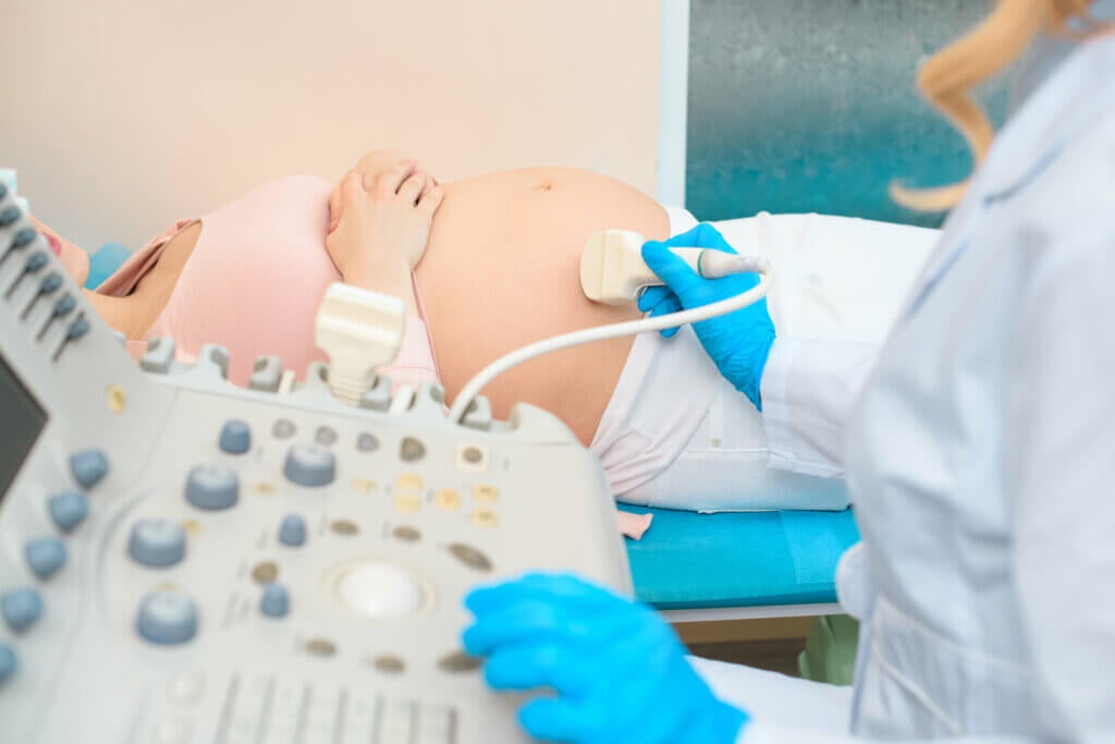
Once the pregnancy has been confirmed and has begun its course, it is time to undergo an ultrasound. This imaging technique is based on ultrasound, which means that it generates two-dimensional or three-dimensional images from sound waves. It’s recommended to carry out this test in week 12, although under prescription it can be advanced.
To carry out this technique, a probe called a transducer is used that is positioned directly on the abdomen of the patient after the application of a gel, which is the main conductor of ultrasound. Once the mother is positioned, high-frequency sound waves are emitted from the transducer, travel through the gel, and enter the maternal environment.
The transducer picks up the sounds that bounce off the body, and a computer uses the information to generate images of the inside of the fetal environment. As the picture generated is in real-time, the fetal movements and the organs near it can also be observed live. This test is done during the first trimester with the following goals in mind:
- To confirm the normality of the pregnancy.
- To learn more about the advancement of the fetus and its development.
- To determine certain complications: here we can detect ectopic pregnancies (if the fetus develops outside the uterine womb) and possible threats of miscarriage.
- To know the fetal heart rate. Basal values range from 120 to 160 beats per minute. Any value above the upper limit is considered a tachycardia.
- To detect multiple pregnancies: If there are twins, twins, or triplets, for example. Abnormally high hGC concentrations in the maternal body can also indicate this event, but ultrasound confirms this.
- Detect problems in the mother’s placenta, uterus, and pelvic environment.
The test lasts 30 to 60 minutes and is not associated with any complications. However, it should be noted that ultrasound doesn’t allow us to see fetal problems with absolute accuracy during the first trimester. In the event of any suspicion or abnormality, specific tests will be required.
In early pregnancy, the ultrasound probe can be placed directly into the vagina.
2.1 Nuchal translucency examination (11-14 weeks)
Many sources state that the nuchal translucency exam is different than the typical ultrasound. In any case, and as indicated by the Natalben portal, it’s an extra test that is part of the general picture collected during the first trimester ultrasound.
The nuchal translucency test tries to measure the thickening of the fetal nape crease (a section at the back of the neck). All unborn babies have some fluid in this area, but those who will develop Down syndrome or other genetic variations accumulate something more than normal.
The procedure is the same as in the previous case (ultrasound), but it should be noted that this time the probe has to be placed in the abdominal environment and not in the vaginal area. The mother may be advised to drink some fluid 2-3 hours before the test, as this favors the filling of the bladder and the transmission of the tubes in the uterine environment.
Normal results of one of these tests or tests during pregnancy are as follows:
- At 11 weeks: up to 2 millimeters
- At 13 weeks and 6 days: up to 2.8 millimeters
The fact that a fetus has a slightly higher than normal nuchal translucency value does not always indicate a genetic abnormality. In any case, this parameter has been associated with Down syndrome, trisomy 18, trisomy 13, and Turner syndrome, among other imbalances of this type.
3. Blood and urine tests
The blood test is the early diagnostic support for almost any condition not visible to the naked eye. Therefore, it’s essential that the mother undergoes one of these tests several times during pregnancy, the first being recommended at 9-13 weeks after confirmation.
Preparing for a blood test is very easy. You just have to go to the designated health center on an empty stomach first thing in the morning and wait for the blood to be drawn. Some people prefer to lie down during the extraction process (which takes no more than 1-2 minutes) for fear of needles, but this is the biggest complication.
Parameters quantified in these pregnancy tests and analyses
Some of the parameters that are monitored with this analysis are the following:
- Complete blood count: The complete blood count is used to infer the general health of the patient. This reports very relevant information on the number, composition and proportions of the elements in the blood (erythrocytes, hematocrit, hemoglobin, leukocyte formula, and the number of platelets).
- Blood glucose analysis: The amount of glucose in the blood allows a doctor to detect gestational diabetes, a pathology that affects 1-14% of pregnant women. During pregnancy, fasting blood glucose should not exceed 95 mg / dL, one hour after eating it is less than 140 mg / dL and 2 hours later it is less than 120 mg / dL.
- Determination of blood group and Rh: We will focus more on this issue in the next section.
- Indirect Coombs test: Again, we will focus more on this in future sections.
- Antibody analysis: This allows us to detect the presence of circulating antibodies for diseases such as HIV, viral hepatitis, rubella or syphilis. It seeks to diagnose those pathologies that can be transmitted to the fetus or indirectly damage its development.
- Creatinine in the blood: An estimated measure of the general functioning of the kidneys. Normal blood creatinine levels for a pregnant woman are normally between 0.4-0.8 mg / dL.
It should be noted that this test is usually accompanied by a urinalysis. This is done to quantify the amount of chorionic gonadotropin expelled during urination. It also serves as a support method for blood analysis and detects signs of infection, sugar levels, and protein levels in the urine.
Let’s take a closer look at some of the mentioned parameters.
3.1 Determination of the Rh factor
As indicated by the Health Library portal, the Rh test is used to find out if the mother has a specific protein (Rh factor) on the surface of her red blood cells. These types of pregnancy tests and analyses are very important, since they quantify the probabilities of a fetal-maternal incompatibility.
If a mother is Rh negative and the fetus is Rh positive (inherited from the father), the pregnancy is in some danger. When the unborn’s red blood cells come into contact with the maternal blood system, the pregnant woman’s immune system will recognize her child’s blood as harmful and pathogenic. Then, it will create anti-Rh antibodies that will cross the placenta.
Rh incompatibility can occur by 2 main mechanisms:
- The fetus’ blood enters the mother’s bloodstream, either from an overly invasive obstetric procedure, from a miscarriage, from certain traumas, or from a normal delivery. The maternal immune system recognizes the fetus as harmful and tries to eliminate it.
- An Rh-negative mother receives a transfusion from an Rh-positive person: if this happens (which it should not, as it would cause medical problems). When the baby begins to gestate the mother will already have anti-Rh antibodies.
Rh incompatibility can lead to miscarriage, anemia, and problems in future pregnancies. This test is essential to prevent mishaps in the current pregnancy and in others that may take place at other times.
3.2 Indirect Coombs test
The indirect Coombs test is closely related to the previous section. In this case, it isn’t quantified if the mother and fetus are Rh negative or positive: only the specific antibodies are looked for in the mother’s blood. The results are interpreted as follows, as indicated by the Cigna portal:
- Normal (negative) result: The mother hasn’t yet produced specific antibodies against fetal blood. This means that the fetus is not in danger.
- Abnormal result (positive): The mother does have circulating anti-Rh antibodies, that is, she is sensitized. This doesn’t indicate that the fetus has been damaged, but rather that there is some possibility of it occurring.
As you can see, a mother being Rh negative and an Rh positive child doesn’t always predict a miscarriage. For this to occur, there must be an exchange of blood from the fetus to the pregnant woman and a considerable increase in antibodies, a situation that can be avoided with proper medical control.
4. Other prenatal diagnostic techniques

So far, we have presented you with 3 of the tests and analyses that are carried out during pregnancy (the most important ones) and some of their peculiarities, but there are many more. In the following lines, we would like to show you some specific tests that actively look for anatomical and physiological abnormalities in the fetal environment.
Prenatal diagnostic techniques look for abnormalities in the fetal neural tube, chromosomal mismatches (in the karyotype), and genetic mutations, among many other alterations. Some are more invasive than others and can cause spontaneous miscarriages.
Specific diagnostic techniques
Some of the most important are the following:
- Fetal cells in maternal blood (CFSM): As its name suggests, this test is based on detecting the number of cells from the fetus that have infiltrated the pregnant woman’s bloodstream. These cell bodies hold all of the baby’s genetic information, so they can be used for future diagnoses.
- Preimplantation genetic diagnosis (PGD): These tests are used in in vitro fertilization conditions before implanting future embryos in the mother’s uterus. Although it may seem cold, selecting the most viable pre-embryos will prevent future abortions in the mother.
- Collection of transcervical trophoblast cells: This allows us to detect the biological gender of the fetus before its time and identify aneuploidies (an abnormal number of chromosomes in any of its pairs).
- Chorionic villus sampling: A sample of the chorionic villus (portion of the fetal membrane that gradually forms the fetal side of the placenta) is obtained in order to glimpse the DNA, karyotype, and fetal enzyme activity.
All these tests are aimed at anticipating the need for medical or surgical treatment, giving the parents the opportunity to abort (if they wish), or psychologically preparing the parents that something is probably wrong during pregnancy. Where it is allowed, couples can decide whether or not they want to continue with a pregnancy.
5. Amniocentesis
There are many more types of tests and tests that can be performed during pregnancy. However, we would like to finish this article talking about amniocentesis. As indicated by the Mayo Clinic, in this procedure a certain amount of amniotic fluid is extracted from the uterus (20 milliliters), which contains fetal cells and various proteins of clinical interest.
Amniocentesis is carried out for these reasons, among many others:
- Diagnosis of a fetal infection: The term intra-amniotic infection refers to the presence of harmful pathogens in the tissues around the fetus, including the amniotic fluid, the placenta, and the fetal membranes.
- Fetal anemia: As we have said, Rh incompatibility can cause anemia in the fetus. Amniocentesis helps detect this condition.
- Genetic testing: Fetal cells floating in amniotic fluid can be collected in order to detect genetic problems. However, the collection of fetal cells in maternal blood is less invasive. Amniocentesis is carried out when something is already suspected to be wrong.
- Treatment of polyhydramnios: Sometimes the mother can accumulate too much amniotic fluid in pregnancy. This technique helps alleviate symptoms.
- Paternity analysis: The collection of the fetus’ DNA allows it to be compared with the parental ones. In this way, it is confirmed who the father and mother are before birth.
Amniocentesis is one of the most invasive pregnancy tests and reports a miscarriage risk of 0.06% (1 in 1600). In addition, it can only be performed from the 15th week of pregnancy, because before this moment enough amniotic fluid has not been produced. Therefore, its use is reserved to cases where it is strictly necessary.
The variety of tests and analyses during pregnancy
There are many, many tests that can be carried out during pregnancy. Ultrasound, blood tests, and urinalysis are the most common, while amniocentesis or chorionic villus sampling (as examples) are reserved for special cases. These last techniques are very invasive.
Not all tests are carried out once. For example, an average of 3 ultrasounds and 3 blood tests are required during pregnancy (one per trimester). Only in this way is it possible to monitor the fetus with maximum efficiency and avoid problems, no matter how slight.
- 4 Common Pregnancy Complications, Johns Hopkins medicine. Recogido a 5 de octubre en https://www.hopkinsmedicine.org/health/conditions-and-diseases/staying-healthy-during-pregnancy/4-common-pregnancy-complications
- Ultrasonido general, Radiology.info. Recogido a 5 de noviembre en https://www.radiologyinfo.org/es/info/genus#:~:text=El%20ultrasonido%20es%20un%20examen,se%20las%20conoce%20como%20ecograf%C3%ADa.
- La ecografía de la translucencia nucal, Natalben. Recogido a 5 de noviembre en https://www.natalben.com/guia-embarazo/embarazo-tercer-mes/ecografia-primer-trimestre
- Determinación del grupo Rh, Health Library. Recogido a 5 de noviembre en https://healthlibrary.brighamandwomens.org/Spanish/RelatedItems/167,Rh_typing_ES
- Prueba de Coombs indirecta, Cigna. Recogido a 5 de noviembre en https://www.cigna.com/es-us/individuals-families/health-wellness/hw/prueba-de-coombs-indirecta-hw139013
- Amniocentesis, Clínica Mayo. Recogido a 5 de noviembre en https://www.mayoclinic.org/es-es/tests-procedures/amniocentesis/about/pac-20392914