All You Need to Know About Ultrasound
Ultrasound is a diagnostic imaging technique. It uses high-frequency sound waves to look at organs and other elements inside the body. It’s mainly used to observe the heart, blood vessels, kidneys, the liver, and joints.
In addition to this, it’s the first choice technique to monitor pregnancies and observe the fetus. Unlike X-rays, ultrasound doesn’t expose the patient to radiation, so it’s safer.
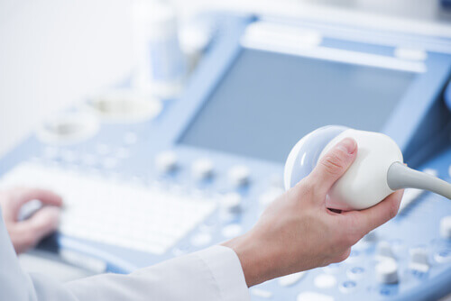
A small instrument called a transducer, very similar to a ‘microphone’, emits the ultrasound waves. These waves are then transmitted to the area of the body being studied and their echo is received.
The transducer picks up the echo from the sound waves, and a computer converts the echo from the waves into images that appear on the screen.
Knowing the origins of ultrasound
In 1942, an Austrian psychiatrist named Karl Dussik was trying to detect brain tumors by recording the passage of the sound beam through the skull of patients. With this technique, he tried to identify the ventricles by measuring the attenuation of the ultrasound through the skull, which he called hyperphonography of the brain.
With this procedure, by means of an oscillograph, a device that records sound waves or undulations, one-dimensional records were obtained. They are traces similar to those of an electrocardiogram or an encephalogram. From that time on, many more sound and image-based diagnostic methods were invented and discovered.

It was in the 1980s, with the incorporation of the computer in ultrasound machines, when the procedure was revolutionized. With this invention, it was possible to process the waves directly and obtain images in real-time. It was also now possible to print the images that the specialist considered important and to find out the measurements of the organs with total precision.
Nowadays, ultrasound can obtain three-dimensional images and others in four dimensions that give a real close-up of the image, especially in fetuses.
What materials are needed to do an ultrasound?
Ultrasound is an easy and safe technique to perform. The machinery is made up of two parts and, in order for it to work, it requires the application of a special gel. Therefore, carrying out an ultrasound requires:
- Gel: This is applied to the area to be studied and acts as a conductor of ultrasound.
- Echograph and transducer: This sends the ultrasound waves that are reflected in the internal body structures. This information is analyzed by the computer, which creates an image of the organ being scanned on the screen. There are different types of ultrasound, depending on the structure to be examined: linear, sectorial, convex, and endocavitary.
Also read: Blood Clots: Whar Are the Warning Signs?
Types of ultrasounds
The most commonly used types of ultrasound are the following:
- Abdominal: This is used to detect any abnormality of the abdominal organs, such as the kidneys, liver or pancreas. You can see stones in the gallbladder or tumors, for example.
- Renal: The kidneys and urinary tract are examined.
- Obstetric: This type of ultrasound is used to monitor the development of the fetus during pregnancy.
- Pelvic: Thanks to this technique, the cause of pelvic pain, such as an ectopic pregnancy in women, can be found. Tumors or masses can also be detected.
- Breasts: Lumps in the breast tissue can be examined.
- Scrotum: This is used to further investigate testicular pain.
- Locomotor: Any nodule noted during a physical examination can be studied.
- Others: vascular, echocardiogram, interventional, thyroid, prostate, etc.

You may be interested in: What Is MR Enterography?
New ultrasound techniques
A traditional ultrasound modality that is being used more and more is Doppler ultrasound. Its name comes from the Doppler effect that it uses. It consists of an apparent change in frequency in a wave.
It’s used, above all, as a first step in the study of blood vessels. However, in many cases, an arteriography or venography is also usually required.
On the other hand, we must mention another variety of traditional ultrasound, 4-dimensional or 4D ultrasound. This technique is used specifically in the gestational process, as the image is somewhat sharper.
Thanks to this technique, it’s possible to clearly perceive the size of the baby and its movements, in addition to knowing its sex with precision. However, it must be taken into account that this modality doesn’t imply any advances in the diagnosis with respect to 3D ultrasound.
- Paola Paolinelli, D. G. (2004). Ecografía Doppler: Principios y aplicaciones. Abril.
- Pascual, M. Á., Hereter, L., Graupera, B., Cid, M. F., & Dexeus, S. (2006). Ecografía 3D/4D en ginecología: Técnica y metodología. Progresos En Obstetricia y Ginecologia. https://doi.org/10.1016/S0304-5013(06)72605-X
- Segui, P., & Espejo, S. (2010). Ecografia. In Imaging diagnostico. https://doi.org/10.1007/978-88-470-1510-4_10
- Díaz-Rodríguez, N., Garrido-Chamorro, R. P., & Castellano-Alarcón, J. (2009). Ecografía: principios físicos, ecógrafos y lenguaje ecográfico. SEMERGEN – Medicina de Familia. https://doi.org/10.1016/s1138-3593(07)73916-3