The Stages of Breast Cancer
The diagnosis of a malignant lump in the breast is shocking news, both for those affected and for their loved ones. The prognosis and therapeutic options will depend on the characteristics of the condition. In this sense, there are different stages of breast cancer, which are related to its extension or evolution.
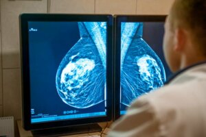
The classification of stages can be both clinical and pathologic. The clinical form is made based on the different imaging studies and biopsies performed. For its part, pathological determination is only possible after surgery, in which a specialist will analyze the extracted tissue.
Breast cancer staging system

The different stages of breast cancer are determined using the TNM system. This model evaluates 3 fundamental characteristics, which are the size of the tumor, the extension to the nodes, and the metastasis. Each of these characteristics has a precise classification, which will be combined to form the specific stage classification.
In recent years, the presence of certain markers such as estrogen and progesterone receptors has also been evaluated, as well as the production of HER2 protein. Studies have revealed that these results can change the prognosis of patients in up to 40% of cases, so it’s important not to overlook them.
Tumor size (T)
Knowing the size of the primary tumor in breast cancer is essential, since it not only influences the symptoms presented, but also guides the possible anatomical changes presented. This category is divided into numbers from 0 to 4, which help describe the neoplasm more precisely. The subdivisions of tumor size are as follows:
- Tx: This is a primary tumor that cannot be evaluated, either due to its small size or its location.
- T0: This means that there is no evidence to indicate the presence of breast cancer in the evaluated breast.
- Tis: This subdivision is used to indicate that there is a carcinoma in situ, either ductal or lobular.
- T1: This indicates the presence of a malignant mass that measures less than 2 centimeters (0.8 inches)
- T2: This is used for a tumor that measures more than 2 centimeters but less than 5 centimeters (0.8 to 2 inches).
- T3: This is used to describe the presence of any malignant mass larger than 5 centimeters (2 inches).
- T4: This subdivision is used to establish that the tumor has affected the chest wall, the skin, or that it’s an inflammatory breast cancer.
Lymph nodes (N)
The letter N on the TNM scale refers to the spread of cancer cells to the lymph nodes. The specialist must consider the affection of the nodes located in the axillary region, below the sternum, and those near the clavicle.
The study of the affected nodes is carried out through the observation of the nodes under a microscope, either after a biopsy or after a surgical removal as part of the treatment of the disease. It’s important to define the number of affected nodes and their location, and we can find the following subdivisions:
- Nx: This indicates that the lymph nodes couldn’t be evaluated, in most cases due to a previous removal.
- N0: This can mean 2 things, either that there is no involvement of the nearby lymph nodes or that they have an invasion of less than 0.2 millimeters.
- N1: This is used to indicate that the disease has affected between 1 and 3 nodes located in the axillary region or behind the sternum.
- N2: In this case the number of affected axillary or sternal nodes varies between 4 and 9.
- N3: This subdivision is used when there are 10 or more axillary or sternal nodes affected. It is also used when the nodes located above or below the clavicle are involved.
Metastasis (M)
Metastasis indicates that the cancer has already spread to other parts of the body, so it is no longer considered early or local-stage cancer. This category has only 3 subdivisions, which are the following:
- Mx: When metastasis to other organs cannot be assessed.
- M0: There is no evidence of spread to other parts of the body or the size of the spread is less than 0.2 millimeters.
- M1: There is metastasis in other tissues, that is, cancer cells have begun to grow in other areas.
Stages of breast cancer
Generally speaking, there are 5 different stages for breast cancer that are listed from 0 to 4 in Roman numerals. They are established through the combination of the sub-classifications of the TNM system. As the numbering progresses, the more serious the disease will be.
Stage 0
A cancer is said to be stage 0 when it has a Tis, N0, and M0 classification. In this sense, it is said that there is the presence of a carcinoma in situ, either ductal or lobular. Cancer cells are present, however, they haven’t initiated the invasion into the surrounding tissue.
This is one of the initial stages of breast cancer, so the prognosis is good according to different studies. Conservative surgical treatment has shown good results, however, radical mastectomy is necessary in cases of large tumors.
Stage I
Stage I is another stage that indicates that the cancer hasn’t yet advanced. It has begun to invade the surrounding tissue, but it is still limited to the breast and is small. This stage can be divided into 2 different categories, which are the following:
- Ia: This is a tumor classified as T1, N0, M0, so it’s smaller than 2 centimeters (0.8 inches) and it doesn’t have lymphatic spread or metastasis.
- Ib: This is a T0 or T1, N1, Mo tumor, which means that it has spread to less than 3 axillary or sternal lymph nodes with an affection of smaller than 2 millimeters, with or without a primary tumor.
During these stages, one of the preferred diagnostic methods are imaging tests such as mammography and a breast eco-sonogram. The prognosis is also usually good in most cases, depending on the presence of hormone receptors and the production of HER2 protein.
Stage II

This stage is characterized by the presence of a tumor of considerable size, reaching an average of more than 5 centimeters (2 inches). In some cases, the cancer is limited to the breast, but it can affect up to 3 lymph nodes. This stadium also has 2 subdivisions, which are the following:
- IIa: There are two different possibilities. The first is that the tumor is similar to stage Ib, but that the lymph node involvement has an extension greater than 2 millimeters. For its part, the second refers to a T2, N0, M0 tumor, that is, a tumor between 2 and 5 centimeters (0.8 to 2 inches) without lymph node extension.
- IIb: This is a T2, N1, M0 tumor, that is, with a size between 2 and 5 centimeters (0.8 to 2 inches) and with an extension of up to a maximum of 3 lymph nodes. A T3, N0, and M0 tumor can also be treated, which means that it is larger than 5 centimeters (2 inches) in size without lymph node involvement or metastasis.
The large size of tumors in this category makes them easier to detect with breast self-examination. The cancer has started to spread at this stage, so it’s necessary to start treatment as soon as possible in order to prevent the disease from progressing.
Stage III
This is one of the most advanced stages of breast cancer, just before it begins to metastasize to distant organs. At this point in the disease, a large tumor, extensive lymphatic spread, and involvement of adjacent tissue, such as the chest wall and skin, may be found. It is divided into 3 subcategories depending on the severity:
- IIIa: This is a tumor of any size that has affected between 4 and 9 lymph nodes, making it a T0, T1 or T2, N2, M0. It can also be a tumor greater than 5 centimeters (2 inches) with an extension to a maximum of 3 nodes, that is, a T3, N1, and M0 neoplasm according to the TNM scale.
- IIIb: This is a tumor of any size that has affected the chest wall, generated an ulceration on the skin, or became an inflammatory cancer. In addition, there may be involvement of up to 9 lymph nodes, which is why it’s classified according to the TNM as T4, N0, N1 or N2, M0.
- IIIc: This is a neoplasm of any size that has managed to spread to 10 lymph nodes or more, yet has not yet metastasized. In this sense, it is classified according to the TNM with any T, N3, M0.
Stage IV
Of all the stages of breast cancer, this is the most advanced, and, therefore, the one with the worst prognosis. In stage IV the disease has spread beyond the breast, affecting other organs. Unfortunately, studies show there is no definitive cure, although there are different treatments that improve the quality of life.
Among the organs most frequently affected by metastatic cancer are the bones and liver. Many types of cancer are likely to metastasize, so proper monitoring and control is important.
Treatment is important at all stages
The stages of breast cancer are directly related to the progression of the disease and its prognosis. In this sense, the most advanced stages have a very poor prognosis, so it’s important to prevent the disease from reaching such an extreme.
There are many therapeutic options that can be the definitive cure in the initial stages of the disease, highlighting the importance of a timely diagnosis. It’s always advisable to have constant medical check-ups and a monthly breast self-examination. In addition, you should see a doctor in the presence of any strange symptoms.
The diagnosis of a malignant lump in the breast is shocking news, both for those affected and for their loved ones. The prognosis and therapeutic options will depend on the characteristics of the condition. In this sense, there are different stages of breast cancer, which are related to its extension or evolution.
The classification of stages can be both clinical and pathologic. The clinical form is made based on the different imaging studies and biopsies performed. For its part, pathological determination is only possible after surgery, in which a specialist will analyze the extracted tissue.
Breast cancer staging system
The different stages of breast cancer are determined using the TNM system. This model evaluates 3 fundamental characteristics, which are the size of the tumor, the extension to the nodes, and the metastasis. Each of these characteristics has a precise classification, which will be combined to form the specific stage classification.
In recent years, the presence of certain markers such as estrogen and progesterone receptors has also been evaluated, as well as the production of HER2 protein. Studies have revealed that these results can change the prognosis of patients in up to 40% of cases, so it’s important not to overlook them.
Tumor size (T)
Knowing the size of the primary tumor in breast cancer is essential, since it not only influences the symptoms presented, but also guides the possible anatomical changes presented. This category is divided into numbers from 0 to 4, which help describe the neoplasm more precisely. The subdivisions of tumor size are as follows:
- Tx: This is a primary tumor that cannot be evaluated, either due to its small size or its location.
- T0: This means that there is no evidence to indicate the presence of breast cancer in the evaluated breast.
- Tis: This subdivision is used to indicate that there is a carcinoma in situ, either ductal or lobular.
- T1: This indicates the presence of a malignant mass that measures less than 2 centimeters (0.8 inches)
- T2: This is used for a tumor that measures more than 2 centimeters but less than 5 centimeters (0.8 to 2 inches).
- T3: This is used to describe the presence of any malignant mass larger than 5 centimeters (2 inches).
- T4: This subdivision is used to establish that the tumor has affected the chest wall, the skin, or that it’s an inflammatory breast cancer.
Lymph nodes (N)
The letter N on the TNM scale refers to the spread of cancer cells to the lymph nodes. The specialist must consider the affection of the nodes located in the axillary region, below the sternum, and those near the clavicle.
The study of the affected nodes is carried out through the observation of the nodes under a microscope, either after a biopsy or after a surgical removal as part of the treatment of the disease. It’s important to define the number of affected nodes and their location, and we can find the following subdivisions:
- Nx: This indicates that the lymph nodes couldn’t be evaluated, in most cases due to a previous removal.
- N0: This can mean 2 things, either that there is no involvement of the nearby lymph nodes or that they have an invasion of less than 0.2 millimeters.
- N1: This is used to indicate that the disease has affected between 1 and 3 nodes located in the axillary region or behind the sternum.
- N2: In this case the number of affected axillary or sternal nodes varies between 4 and 9.
- N3: This subdivision is used when there are 10 or more axillary or sternal nodes affected. It is also used when the nodes located above or below the clavicle are involved.
Metastasis (M)
Metastasis indicates that the cancer has already spread to other parts of the body, so it is no longer considered early or local-stage cancer. This category has only 3 subdivisions, which are the following:
- Mx: When metastasis to other organs cannot be assessed.
- M0: There is no evidence of spread to other parts of the body or the size of the spread is less than 0.2 millimeters.
- M1: There is metastasis in other tissues, that is, cancer cells have begun to grow in other areas.
Stages of breast cancer
Generally speaking, there are 5 different stages for breast cancer that are listed from 0 to 4 in Roman numerals. They are established through the combination of the sub-classifications of the TNM system. As the numbering progresses, the more serious the disease will be.
Stage 0
A cancer is said to be stage 0 when it has a Tis, N0, and M0 classification. In this sense, it is said that there is the presence of a carcinoma in situ, either ductal or lobular. Cancer cells are present, however, they haven’t initiated the invasion into the surrounding tissue.
This is one of the initial stages of breast cancer, so the prognosis is good according to different studies. Conservative surgical treatment has shown good results, however, radical mastectomy is necessary in cases of large tumors.
Stage I
Stage I is another stage that indicates that the cancer hasn’t yet advanced. It has begun to invade the surrounding tissue, but it is still limited to the breast and is small. This stage can be divided into 2 different categories, which are the following:
- Ia: This is a tumor classified as T1, N0, M0, so it’s smaller than 2 centimeters (0.8 inches) and it doesn’t have lymphatic spread or metastasis.
- Ib: This is a T0 or T1, N1, Mo tumor, which means that it has spread to less than 3 axillary or sternal lymph nodes with an affection of smaller than 2 millimeters, with or without a primary tumor.
During these stages, one of the preferred diagnostic methods are imaging tests such as mammography and a breast eco-sonogram. The prognosis is also usually good in most cases, depending on the presence of hormone receptors and the production of HER2 protein.
Stage II
This stage is characterized by the presence of a tumor of considerable size, reaching an average of more than 5 centimeters (2 inches). In some cases, the cancer is limited to the breast, but it can affect up to 3 lymph nodes. This stadium also has 2 subdivisions, which are the following:
- IIa: There are two different possibilities. The first is that the tumor is similar to stage Ib, but that the lymph node involvement has an extension greater than 2 millimeters. For its part, the second refers to a T2, N0, M0 tumor, that is, a tumor between 2 and 5 centimeters (0.8 to 2 inches) without lymph node extension.
- IIb: This is a T2, N1, M0 tumor, that is, with a size between 2 and 5 centimeters (0.8 to 2 inches) and with an extension of up to a maximum of 3 lymph nodes. A T3, N0, and M0 tumor can also be treated, which means that it is larger than 5 centimeters (2 inches) in size without lymph node involvement or metastasis.
The large size of tumors in this category makes them easier to detect with breast self-examination. The cancer has started to spread at this stage, so it’s necessary to start treatment as soon as possible in order to prevent the disease from progressing.
Stage III
This is one of the most advanced stages of breast cancer, just before it begins to metastasize to distant organs. At this point in the disease, a large tumor, extensive lymphatic spread, and involvement of adjacent tissue, such as the chest wall and skin, may be found. It is divided into 3 subcategories depending on the severity:
- IIIa: This is a tumor of any size that has affected between 4 and 9 lymph nodes, making it a T0, T1 or T2, N2, M0. It can also be a tumor greater than 5 centimeters (2 inches) with an extension to a maximum of 3 nodes, that is, a T3, N1, and M0 neoplasm according to the TNM scale.
- IIIb: This is a tumor of any size that has affected the chest wall, generated an ulceration on the skin, or became an inflammatory cancer. In addition, there may be involvement of up to 9 lymph nodes, which is why it’s classified according to the TNM as T4, N0, N1 or N2, M0.
- IIIc: This is a neoplasm of any size that has managed to spread to 10 lymph nodes or more, yet has not yet metastasized. In this sense, it is classified according to the TNM with any T, N3, M0.
Stage IV
Of all the stages of breast cancer, this is the most advanced, and, therefore, the one with the worst prognosis. In stage IV the disease has spread beyond the breast, affecting other organs. Unfortunately, studies show there is no definitive cure, although there are different treatments that improve the quality of life.
Among the organs most frequently affected by metastatic cancer are the bones and liver. Many types of cancer are likely to metastasize, so proper monitoring and control is important.
Treatment is important at all stages
The stages of breast cancer are directly related to the progression of the disease and its prognosis. In this sense, the most advanced stages have a very poor prognosis, so it’s important to prevent the disease from reaching such an extreme.
There are many therapeutic options that can be the definitive cure in the initial stages of the disease, highlighting the importance of a timely diagnosis. It’s always advisable to have constant medical check-ups and a monthly breast self-examination. In addition, you should see a doctor in the presence of any strange symptoms.
- Hortobagyi GN, Edge SB, Giuliano A. New and Important Changes in the TNM Staging System for Breast Cancer. Am Soc Clin Oncol Educ Book. 2018;38:457-467.
- Cutuli B. Ductal carcinoma in situ in 2019: Diagnosis, treatment, prognosis. Presse Med. 2019;48(10):1112-1122.
- Peart O. Metastatic Breast Cancer. Radiol Technol. 2017 May;88(5):519M-539M.
- Donepudi MS, Kondapalli K, Amos SJ, Venkanteshan P. Breast cancer statistics and markers. J Cancer Res Ther. 2014;10(3):506-11.
- Rakha EA, Reis-Filho JS, Baehner F, Dabbs DJ et al. Breast cancer prognostic classification in the molecular era: the role of histological grade. Breast Cancer Res. 2010;12(4):207.
- Li J, Chen Z, Su K, Zeng J. Clinicopathological classification and traditional prognostic indicators of breast cancer. Int J Clin Exp Pathol. 2015;8(7):8500-5.
Este texto se ofrece únicamente con propósitos informativos y no reemplaza la consulta con un profesional. Ante dudas, consulta a tu especialista.


