The 4 Most Common Tongue Diseases or Conditions

Escrito y verificado por la odontóloga Vanesa Evangelina Buffa
The most common tongue diseases may seem trivial or unimportant, but it’s necessary to pay close attention to them. These are conditions that are usually benign, but that could also hide symptoms of more serious pathologies.
Sometimes, the first consultation before the signs that are evident is specified with a general practitioner. However, the best thing would be to go and see a dentist. Oral health professionals are trained to identify, diagnose, and treat conditions of the tongue.
Possibly, canker sores are the ones that top the list of most common tongue diseases. However, we’ll also talk about oral lichen planus, trauma, and infections.
The most common tongue diseases
1. Canker sores
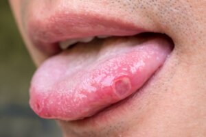
Canker sores on the tongue are very painful. Anyone who has had one will know what we are talking about. They bother us when we’re eating and talking, and even swallowing saliva.
They are small, rounded-looking ulcerations that can appear on any soft tissue in the mouth. Therefore, we are susceptible to developing on the tongue.
They may have a yellowish color at first, and then turn white. They’re surrounded by an intense red halo.
Sometimes there’s a single canker sore on the tongue (when we bite down while chewing). Other times a group develops that may or may not be close to each other; they can also join.
The causes of canker sores are discussed in the scientific field. Although it’s known that traumas can cause its appearance, there’s no certainty about which substances are linked to the disorder. Some people are more susceptible to a certain type of food or an ingredient, for example. Stress must also be considered as a trigger for them.
Canker sores don’t require treatment because they go away on their own within a week. However, a doctor may prescribe painkillers or local anesthetics to help you cope with the discomfort.
Larger canker sores
There’s another situation with larger canker sores, that is, those that are more than 1 centimeter (0.4 inches) in diameter and that tend to heal over a week. Around 20% of cases of canker sores fall within this category.
The healing time is longer in these cases and can be as long as 4 weeks, which complicates healing and increases the possibility of leaving a mark inside the mouth.
Therefore, in people who have a recurrence of this condition, a more intensive approach to treatment is preferred, with topical medication and systemic anti-inflammatory drugs if the dentist or doctor considers it necessary.
Two special clinical forms of canker sores are the following:
- Newman’s bipolar aphthosis: Ulcerations that appear simultaneously in the mouth and in the genital region.
- Sutton’s disease: The canker sores are located near a minor salivary gland.
Differential diagnosis of canker sores
Although it may seem that this is one of the most common diseases of the tongue and shouldn’t be too difficult to diagnose, it can easily be confused with other disorders. In fact, it is advisable to carry out a clear and concise anamnesis so as to be sure.
Oncological processes and manifestations of systemic diseases that could give some manifestation on the tongue must be ruled out. One of these problems is inflammatory bowel disease.
On the other hand, several reports have indicated that the COVID-19 infection caused oral symptoms. Among them, canker sores ranked first in prevalence statistics.
2. Oral lichen planus on the tongue

Oral lichen planus is a pathology of the mucous membranes and the skin. It’s chronic and can affect the soft tissues of the mouth, including the tongue. In general, it evolves in acute episodes or outbreaks.
Its origin is autoimmune. By mistake, the body’s defenses attack the epithelial cells. This leads to the characteristic lesions of the disease.
Due to its autoimmune nature, it isn’t uncommon for it to appear in people who have other disorders of their defense system. A link has also been found with patients who have hepatitis C.
Oral lichen planus isn’t contagious, and isn’t transmitted between humans. In addition, its course is chronic, so those affected have to find ways to deal with the symptoms when flare-ups occur.
The most frequent manifestations are the following:
- Raised whitish spots
- Sores or ulcers
- Reddish painful lesions
The mere presence of symptoms may not be sufficient for a definitive diagnosis of lichen planus on the tongue. The dentist may request a biopsy of the lesions to confirm their suspicion. In addition, immunological analyzes can be carried out on the sample to determine some proteins typical of the disorder, which increases the veracity of the diagnosis.
According to the patient’s history and the assessment made by the expert, blood tests may or may not be required. This is done to track any associated diseases that could be more severe. For this reason, tests for hepatitis C and autoimmune pathologies such as systemic lupus erythematosus are usually recommended.
Can oral lichen planus become malignant?
There are sufficient scientific elements to establish that oral lichen planus has malignant potential. According to statistics, about 1% of patients with the disease develop cancer in their mouth. And this happens especially when the lesions settle on the tongue.
There are other risk factors that increase the risk of malignancy:
- Being of the female sex
- Being a smoker
- Drinking alcohol
- Having infections by the fungus Candida spp. in the mouth frequently
- Being infected with HIV or the hepatitis C virus.
Is there any treatment?
There’s no curative treatment for oral lichen planus. What is recommended are medications and dietary hygienic measures to alleviate the discomfort.
Anti-inflammatories are the drugs of choice and can be administered orally, topically, or by injection. There are also active ingredients that stimulate immune function, such as tacrolimus, which give acceptable results.
3. Tongue trauma
Tongue trauma is one of the most common diseases this organ can have, and most of us can verify this. Most of us will have bitten ourselves while eating or burned ourselves with very hot food.
Sometimes there is a small bleeding that stops almost immediately, while other times there is hypersensitivity or an ulcer in the area, like the ones we have already explained.
However, there are also chronic cases of tongue trauma. This is frequent in people with dental malocclusion, which, due to the bad position of their teeth, unconsciously harm their tongues.
In some cases, the trauma leads to chronic ulcers that don’t seem to heal and bleed frequently. Of course, treatment focuses on resolving the malocclusion so that the tongue will stop suffering from these lesions.
Tongue trauma can also be caused by the presence of poorly adapted dental prostheses or orthodontic appliances, especially when braces are used. Constant rubbing with sharp edges or irregular points can easily cause tongue injuries.
Another specific situation is the practice of certain sports that increase the risk of tongue trauma. Among them, it’s worth mentioning the following:
- Karate
- Handball
- Basketball
- Skiing
Tongue trauma and oral cancer
As with oral lichen planus, an association was found between chronic irritation of the oral mucosa and the development of a neoplastic pathology. In particular, it seems that cancer on the edge of the tongue is prone to start after repeated trauma to that area.
Therefore, it’s extremely important to identify any factors that can harm the tongue. A prompt resolution could prevent a potential tumor in the future.
4. Candida infection or tongue candidiasis

Finally, the last of the most common diseases of the tongue is candidiasis. This is an infection that can develop inside the mouth due to colonization by the Candida albicans fungus.
Babies, the elderly, people who wear dentures, and patients with autoimmune pathologies are more susceptible to this fungus. In general, if the person is healthy, then it’s very unusual for there to be symptoms due to the presence of the microorganism.
The characteristic sign is a whitish coating that forms on the tongue. It can also spread to other oral regions. In the areas where there’s no white coating, an inflammation of the tissues with a red color and small bleeding is noted.
It isn’t a serious tongue disease, unless the patient has a very compromised immune system. In this situation, there’s a greater risk of complications; the fungus can spread to the esophagus and cause serious systemic problems.
Most of the time the diagnosis is made just by looking at the lesions. Other times it will be necessary to take a sample of the whitish layer to observe it carefully under a microscope.
Once the fungus is confirmed, the treatment is carried out using nystatin. There are other antifungals that can be used, such as fluconazole.
Be aware of the most common tongue diseases
Beyond these four tongue diseases, there are other conditions and symptoms of the mouth that you shouldn’t ignore. If you have any questions, make an appointment with your trusted doctor or dentist. A timely diagnosis can prevent complications and improve your quality of life.
The most common tongue diseases may seem trivial or unimportant, but it’s necessary to pay close attention to them. These are conditions that are usually benign, but that could also hide symptoms of more serious pathologies.
Sometimes, the first consultation before the signs that are evident is specified with a general practitioner. However, the best thing would be to go and see a dentist. Oral health professionals are trained to identify, diagnose, and treat conditions of the tongue.
Possibly, canker sores are the ones that top the list of most common tongue diseases. However, we’ll also talk about oral lichen planus, trauma, and infections.
The most common tongue diseases
1. Canker sores
Canker sores on the tongue are very painful. Anyone who has had one will know what we are talking about. They bother us when we’re eating and talking, and even swallowing saliva.
They are small, rounded-looking ulcerations that can appear on any soft tissue in the mouth. Therefore, we are susceptible to developing on the tongue.
They may have a yellowish color at first, and then turn white. They’re surrounded by an intense red halo.
Sometimes there’s a single canker sore on the tongue (when we bite down while chewing). Other times a group develops that may or may not be close to each other; they can also join.
The causes of canker sores are discussed in the scientific field. Although it’s known that traumas can cause its appearance, there’s no certainty about which substances are linked to the disorder. Some people are more susceptible to a certain type of food or an ingredient, for example. Stress must also be considered as a trigger for them.
Canker sores don’t require treatment because they go away on their own within a week. However, a doctor may prescribe painkillers or local anesthetics to help you cope with the discomfort.
Larger canker sores
There’s another situation with larger canker sores, that is, those that are more than 1 centimeter (0.4 inches) in diameter and that tend to heal over a week. Around 20% of cases of canker sores fall within this category.
The healing time is longer in these cases and can be as long as 4 weeks, which complicates healing and increases the possibility of leaving a mark inside the mouth.
Therefore, in people who have a recurrence of this condition, a more intensive approach to treatment is preferred, with topical medication and systemic anti-inflammatory drugs if the dentist or doctor considers it necessary.
Two special clinical forms of canker sores are the following:
- Newman’s bipolar aphthosis: Ulcerations that appear simultaneously in the mouth and in the genital region.
- Sutton’s disease: The canker sores are located near a minor salivary gland.
Differential diagnosis of canker sores
Although it may seem that this is one of the most common diseases of the tongue and shouldn’t be too difficult to diagnose, it can easily be confused with other disorders. In fact, it is advisable to carry out a clear and concise anamnesis so as to be sure.
Oncological processes and manifestations of systemic diseases that could give some manifestation on the tongue must be ruled out. One of these problems is inflammatory bowel disease.
On the other hand, several reports have indicated that the COVID-19 infection caused oral symptoms. Among them, canker sores ranked first in prevalence statistics.
2. Oral lichen planus on the tongue
Oral lichen planus is a pathology of the mucous membranes and the skin. It’s chronic and can affect the soft tissues of the mouth, including the tongue. In general, it evolves in acute episodes or outbreaks.
Its origin is autoimmune. By mistake, the body’s defenses attack the epithelial cells. This leads to the characteristic lesions of the disease.
Due to its autoimmune nature, it isn’t uncommon for it to appear in people who have other disorders of their defense system. A link has also been found with patients who have hepatitis C.
Oral lichen planus isn’t contagious, and isn’t transmitted between humans. In addition, its course is chronic, so those affected have to find ways to deal with the symptoms when flare-ups occur.
The most frequent manifestations are the following:
- Raised whitish spots
- Sores or ulcers
- Reddish painful lesions
The mere presence of symptoms may not be sufficient for a definitive diagnosis of lichen planus on the tongue. The dentist may request a biopsy of the lesions to confirm their suspicion. In addition, immunological analyzes can be carried out on the sample to determine some proteins typical of the disorder, which increases the veracity of the diagnosis.
According to the patient’s history and the assessment made by the expert, blood tests may or may not be required. This is done to track any associated diseases that could be more severe. For this reason, tests for hepatitis C and autoimmune pathologies such as systemic lupus erythematosus are usually recommended.
Can oral lichen planus become malignant?
There are sufficient scientific elements to establish that oral lichen planus has malignant potential. According to statistics, about 1% of patients with the disease develop cancer in their mouth. And this happens especially when the lesions settle on the tongue.
There are other risk factors that increase the risk of malignancy:
- Being of the female sex
- Being a smoker
- Drinking alcohol
- Having infections by the fungus Candida spp. in the mouth frequently
- Being infected with HIV or the hepatitis C virus.
Is there any treatment?
There’s no curative treatment for oral lichen planus. What is recommended are medications and dietary hygienic measures to alleviate the discomfort.
Anti-inflammatories are the drugs of choice and can be administered orally, topically, or by injection. There are also active ingredients that stimulate immune function, such as tacrolimus, which give acceptable results.
3. Tongue trauma
Tongue trauma is one of the most common diseases this organ can have, and most of us can verify this. Most of us will have bitten ourselves while eating or burned ourselves with very hot food.
Sometimes there is a small bleeding that stops almost immediately, while other times there is hypersensitivity or an ulcer in the area, like the ones we have already explained.
However, there are also chronic cases of tongue trauma. This is frequent in people with dental malocclusion, which, due to the bad position of their teeth, unconsciously harm their tongues.
In some cases, the trauma leads to chronic ulcers that don’t seem to heal and bleed frequently. Of course, treatment focuses on resolving the malocclusion so that the tongue will stop suffering from these lesions.
Tongue trauma can also be caused by the presence of poorly adapted dental prostheses or orthodontic appliances, especially when braces are used. Constant rubbing with sharp edges or irregular points can easily cause tongue injuries.
Another specific situation is the practice of certain sports that increase the risk of tongue trauma. Among them, it’s worth mentioning the following:
- Karate
- Handball
- Basketball
- Skiing
Tongue trauma and oral cancer
As with oral lichen planus, an association was found between chronic irritation of the oral mucosa and the development of a neoplastic pathology. In particular, it seems that cancer on the edge of the tongue is prone to start after repeated trauma to that area.
Therefore, it’s extremely important to identify any factors that can harm the tongue. A prompt resolution could prevent a potential tumor in the future.
4. Candida infection or tongue candidiasis
Finally, the last of the most common diseases of the tongue is candidiasis. This is an infection that can develop inside the mouth due to colonization by the Candida albicans fungus.
Babies, the elderly, people who wear dentures, and patients with autoimmune pathologies are more susceptible to this fungus. In general, if the person is healthy, then it’s very unusual for there to be symptoms due to the presence of the microorganism.
The characteristic sign is a whitish coating that forms on the tongue. It can also spread to other oral regions. In the areas where there’s no white coating, an inflammation of the tissues with a red color and small bleeding is noted.
It isn’t a serious tongue disease, unless the patient has a very compromised immune system. In this situation, there’s a greater risk of complications; the fungus can spread to the esophagus and cause serious systemic problems.
Most of the time the diagnosis is made just by looking at the lesions. Other times it will be necessary to take a sample of the whitish layer to observe it carefully under a microscope.
Once the fungus is confirmed, the treatment is carried out using nystatin. There are other antifungals that can be used, such as fluconazole.
Be aware of the most common tongue diseases
Beyond these four tongue diseases, there are other conditions and symptoms of the mouth that you shouldn’t ignore. If you have any questions, make an appointment with your trusted doctor or dentist. A timely diagnosis can prevent complications and improve your quality of life.
- Alaizari, N. A., et al. “Hepatitis C virus infections in oral lichen planus: a systematic review and meta‐analysis.” Australian dental journal 61.3 (2016): 282-287.
- Asensi Anta, E., et al. “Factores asociados a la malignización del liquen plano oral. Revisión de la literatura.” Avances en Odontoestomatología 35.3 (2019): 131-137.
- Bombeccari, G., et al. “Large oral ulcer of tongue related to dental trauma.” (2017): 51-54.
- Egg-Merino, Antonella, Vanessa Ortega-Pachas, and Manuel Antonio Mattos-Vela. “¿ Qué repercusiones tiene la infección por COVID-19 en la cavidad bucal?.” Revista Estomatológica Herediana 32.2 (2022): 203-204.
- Gómez, Alexis Siré, Carlos Albornoz López del Castillo, and Yanelys Cabrera Villalobos. “Síndrome de sutton.” Archivo Médico Camagüey 6.5 (2015).
- Ineichen, Jasmin, et al. “Dental trauma and tongue injuries in professional alpine ski racing—A worldwide survey.” Dental traumatology 37.3 (2021): 414-418.
- Jajam Maturana, M., and S. Niklander Ebensperger. “Liquen plano oral: recomendaciones para su diagnóstico y tratamiento.” Avances en Odontoestomatología 38.1 (2022): 30-39.
- Lyu, Xin, et al. “Efficacy of nystatin for the treatment of oral candidiasis: a systematic review and meta-analysis.” Drug design, development and therapy 10 (2016): 1161.
- Millsop, Jillian W., and Nasim Fazel. “Oral candidiasis.” Clinics in dermatology 34.4 (2016): 487-494.
- Otero Rey, E., et al. “Candidiasis oral en el paciente mayor.” Avances en odontoestomatología 31.3 (2015): 135-148.
- Rojas, Maria Elena Pereda, Yamily González Cardona, and Luis Wilfrido Torres Herrera. “Actualización sobre liquen plano bucal.” Correo Científico Médico de Holguín 20.3 (2016): 539-555.
- Singhvi, Hitesh Rajendra, Akshat Malik, and Pankaj Chaturvedi. “The role of chronic mucosal trauma in oral cancer: A review of literature.” Indian Journal of Medical and Paediatric Oncology 38.01 (2017): 44-50.
- Tomaz, Aline, et al. “Potencial de transformación maligna del liquen plano oral: estudio retrospectivo.” International journal of odontostomatology 9.3 (2015): 511-517.
- Vaillant, L., M. Samimi, and D. Parent. “Aftas, aftosis, enfermedad de Behçet.” EMC-Dermatología 50.2 (2016): 1-14.
- Weber, P., and F. Pascal. “Aftas y aftosis.” EMC-Tratado de Medicina 21.4 (2017): 1-7.
- Ybarra, Patricia María, Dematología Médica Homeópata, and Adjunta Fundación Médica Homeopática Vitalis. “ESTOMATITIS y AFTAS: Actualización Clínica y Revisión de Rúbricas Repertoriales.”
Este texto se ofrece únicamente con propósitos informativos y no reemplaza la consulta con un profesional. Ante dudas, consulta a tu especialista.


