How Do You Know if It's Skin Cancer or a Mole?

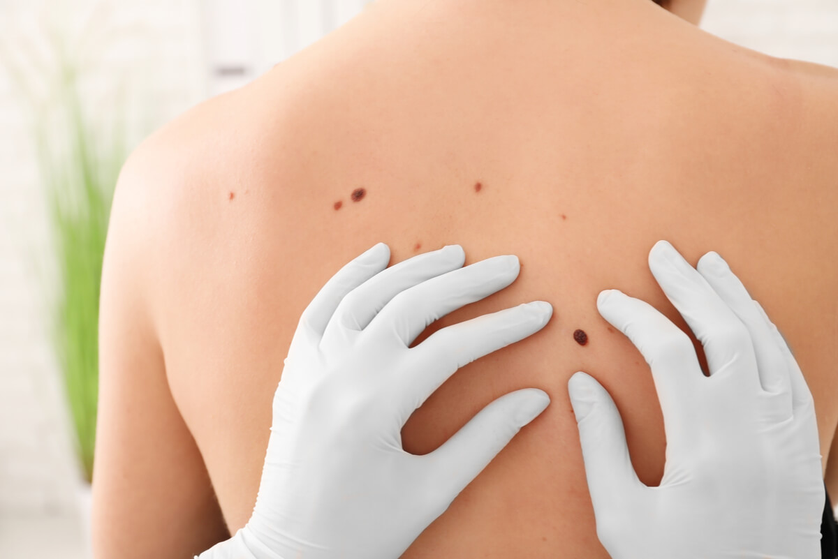
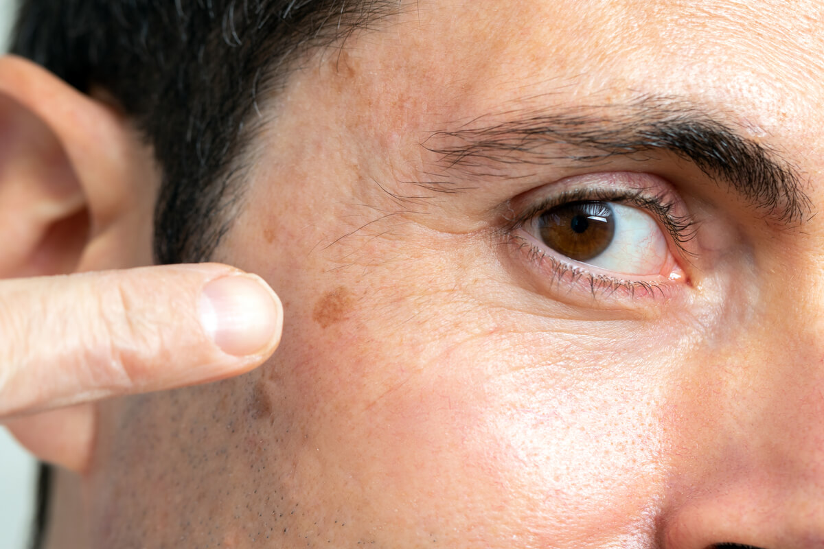
Early detection of skin cancer offers a better prognosis and is associated with a lower morbidity rate. In general, benign moles are often confused with neoplastic pathologies (cancer) and this can alarm affected patients. Are you interested in knowing how to know if a lesion is skin cancer or a mole? Next up, we’ll tell you.
At present, the identification of skin cancer constitutes a great challenge for health professionals. In most cases, these skin lesions can go unnoticed and cannot be differentiated from a common spot or mole. However, there are some characteristics that allow us to suspect if it’s skin cancer or a mole with the naked eye.
Differences between skin cancer and a mole

Moles are defined as ‘benign melanocytic growths that usually appear as small skin-colored or brown spots, papules or nodules’. Studies suggest that they’re only present in 1% of newborns and appear from 6 to 12 months of age. They continue to increase in quantity and size until they’re 25 years old.
In general, it’s rare that a single mole can become malignant and lead to skin cancer. However, the presence of a large number of typical moles or any dysplastic mole should be kept under continuous supervision. Some research claims that the presence of 50 or more moles greatly increases the risk of melanoma.
In the initial identification and differentiation between skin cancer and a mole by the patient, it’s important to consider the details visible to the naked eye. In this sense, the ABCDE rule must be followed to recognize the characteristics of a skin neoplasm. They evaluate the asymmetry, border, color, diameter and elevation of all skin lesions.
Asymmetry
Benign moles can occur anywhere on the body and in a wide variety of ways. They range from a freckle or blemish to a small raised bump on the skin. However, most are characterized by being rounded and maintaining an evident symmetry in both halves.
On the other hand, melanoma-type skin cancer lesions usually appear as irregularly shaped spots or elevations. It’s common for them to be asymmetrical and have a part that’s larger than the rest. In addition, some can be spread on the fabric, imitating the shape of a wine glass stain on a handkerchief.
Border
Regarding the borders, studies estimate that moles are visualized with the naked eye with smooth edges, uniform and separated from the surrounding skin in most cases. However, melanoma lesions often have uneven and irregular borders, which may have notches or a cotton-like appearance along their structure.
This feature should be considered in direct association with the shape and symmetry of any obvious lesions on the skin’s surface. Also, some moles may appear normal at first and acquire strange characteristics over time. In this sense, it’s important to maintain continuous attention to them.
Color
Color is a fundamental factor in the differentiation between skin cancer and a mole. In fact, its importance leads to an in-depth study with a dermoscopy in consultation with the specialist. This examination usually defines the type of injury and provides guidance on the possible malignancy of a mole in most cases.
In moles, it’s common to find shades of brown, pink or similar to the colors of the skin. However, the main aspect that determines its benignity is that the color remains uniform throughout the entire lesion.
On the other hand, spots or moles suggestive of skin cancer usually present several shades that interpose along them. In melanomas, it’s common to find mixtures of brown, black, and tan tones. Some research affirms that the tones of the borders of the lesion are the most important ones in the identification of cutaneous neoplasms.
Diameter

The dimensions of typical moles can vary greatly according to their shape, and the limit established to consider a lesion as benign is 6 millimeters. In this sense, any lesion that’s above these dimensions must suggest some type of skin cancer.
Some patients use the size of a pencil eraser as a reference for the injury. However, melanomas can appear as large spots or moles.
Evolution
Regular monitoring of skin lesions is often the key to early and timely cancer diagnosis. Skin neoplasms usually appear as moles that change their shape, symmetry, borders, and colors over time. Similarly, they can appear spontaneously on healthy skin.
In some cases, the skin lesions tend to have a hard, lumpy, or scaly consistency that increases the suspicion of cancer. In addition, they become larger and thicker, covering a greater extension of the skin surface.
Concomitant symptoms
When evaluating a suspicious mole or spot, other associated symptoms should be taken into account to support diagnosis. The presence of bleeding, purulent discharge and itching usually guides the attending physician. In some patients, sensation disturbances such as numbness and tingling occur.
Early detection makes a difference
Today, it’s very common for people not to know if they have skin cancer or a mole. This fact worsens the evolution of the disease by not being approached in time. Therefore, if you have several moles, a strange lesion or a spot that appears spontaneously, you should go as soon as possible to your trusted doctor.
In the same way, a routine self-examination of the skin is essential, and it’s one of the most important measures to treat skin cancer in its initial stages. In addition, the use of adequate sun protection and follow-up exams with the dermatologist are associated with a lower risk of skin cancer.
Early detection of skin cancer offers a better prognosis and is associated with a lower morbidity rate. In general, benign moles are often confused with neoplastic pathologies (cancer) and this can alarm affected patients. Are you interested in knowing how to know if a lesion is skin cancer or a mole? Next up, we’ll tell you.
At present, the identification of skin cancer constitutes a great challenge for health professionals. In most cases, these skin lesions can go unnoticed and cannot be differentiated from a common spot or mole. However, there are some characteristics that allow us to suspect if it’s skin cancer or a mole with the naked eye.
Differences between skin cancer and a mole

Moles are defined as ‘benign melanocytic growths that usually appear as small skin-colored or brown spots, papules or nodules’. Studies suggest that they’re only present in 1% of newborns and appear from 6 to 12 months of age. They continue to increase in quantity and size until they’re 25 years old.
In general, it’s rare that a single mole can become malignant and lead to skin cancer. However, the presence of a large number of typical moles or any dysplastic mole should be kept under continuous supervision. Some research claims that the presence of 50 or more moles greatly increases the risk of melanoma.
In the initial identification and differentiation between skin cancer and a mole by the patient, it’s important to consider the details visible to the naked eye. In this sense, the ABCDE rule must be followed to recognize the characteristics of a skin neoplasm. They evaluate the asymmetry, border, color, diameter and elevation of all skin lesions.
Asymmetry
Benign moles can occur anywhere on the body and in a wide variety of ways. They range from a freckle or blemish to a small raised bump on the skin. However, most are characterized by being rounded and maintaining an evident symmetry in both halves.
On the other hand, melanoma-type skin cancer lesions usually appear as irregularly shaped spots or elevations. It’s common for them to be asymmetrical and have a part that’s larger than the rest. In addition, some can be spread on the fabric, imitating the shape of a wine glass stain on a handkerchief.
Border
Regarding the borders, studies estimate that moles are visualized with the naked eye with smooth edges, uniform and separated from the surrounding skin in most cases. However, melanoma lesions often have uneven and irregular borders, which may have notches or a cotton-like appearance along their structure.
This feature should be considered in direct association with the shape and symmetry of any obvious lesions on the skin’s surface. Also, some moles may appear normal at first and acquire strange characteristics over time. In this sense, it’s important to maintain continuous attention to them.
Color
Color is a fundamental factor in the differentiation between skin cancer and a mole. In fact, its importance leads to an in-depth study with a dermoscopy in consultation with the specialist. This examination usually defines the type of injury and provides guidance on the possible malignancy of a mole in most cases.
In moles, it’s common to find shades of brown, pink or similar to the colors of the skin. However, the main aspect that determines its benignity is that the color remains uniform throughout the entire lesion.
On the other hand, spots or moles suggestive of skin cancer usually present several shades that interpose along them. In melanomas, it’s common to find mixtures of brown, black, and tan tones. Some research affirms that the tones of the borders of the lesion are the most important ones in the identification of cutaneous neoplasms.
Diameter

The dimensions of typical moles can vary greatly according to their shape, and the limit established to consider a lesion as benign is 6 millimeters. In this sense, any lesion that’s above these dimensions must suggest some type of skin cancer.
Some patients use the size of a pencil eraser as a reference for the injury. However, melanomas can appear as large spots or moles.
Evolution
Regular monitoring of skin lesions is often the key to early and timely cancer diagnosis. Skin neoplasms usually appear as moles that change their shape, symmetry, borders, and colors over time. Similarly, they can appear spontaneously on healthy skin.
In some cases, the skin lesions tend to have a hard, lumpy, or scaly consistency that increases the suspicion of cancer. In addition, they become larger and thicker, covering a greater extension of the skin surface.
Concomitant symptoms
When evaluating a suspicious mole or spot, other associated symptoms should be taken into account to support diagnosis. The presence of bleeding, purulent discharge and itching usually guides the attending physician. In some patients, sensation disturbances such as numbness and tingling occur.
Early detection makes a difference
Today, it’s very common for people not to know if they have skin cancer or a mole. This fact worsens the evolution of the disease by not being approached in time. Therefore, if you have several moles, a strange lesion or a spot that appears spontaneously, you should go as soon as possible to your trusted doctor.
In the same way, a routine self-examination of the skin is essential, and it’s one of the most important measures to treat skin cancer in its initial stages. In addition, the use of adequate sun protection and follow-up exams with the dermatologist are associated with a lower risk of skin cancer.
- Goldstein A, Tucker M. Dysplastic Nevi and Melanoma. Cancer Epidemiol Biomarkers. 2013; 22(4): 528-532.
- Black S, MacDonald-McMillan B, Mallett X et al. The incidence and position of melanocytic nevi for the purposes of forensic image comparison. Int J Legal Med. 2014; 128: 535–543.
- Chen J, Stanley RJ, Moss RH, Van Stoecker W. Colour analysis of skin lesion regions for melanoma discrimination in clinical images. Skin Res Technol. 2003;9(2):94-104.
- Halem M, Karimkhani C. Dermatology of the head and neck: skin cancer and benign skin lesions. Dent Clin North Am. 2012;56(4):771-90.
- Bahnson AB, Kondratuk KE, Anderson SM. Skin Cancer Education in the Rural Salon. S D Med. 2019;72(6):267-271.
- Jones OT, Ranmuthu CKI, Hall PN, Funston G, Walter FM. Recognising Skin Cancer in Primary Care. Adv Ther. 2020;37(1):603-616.
Este texto se ofrece únicamente con propósitos informativos y no reemplaza la consulta con un profesional. Ante dudas, consulta a tu especialista.


