Differences Between a CT and an MRI

Medicine has advanced dramatically in recent decades, thus allowing professionals to observe the internal tissues of the patient without having to resort to surgery. Many diagnostic methods used today are minimally invasive and report a great deal of information about individual status. Do you know the differences between a CT and an MRI?
Although both techniques allow observing the inner tissue conglomerate of the patient, they have a series of fundamental differences. Keep reading because below, we’ll tell you the uses, functional characteristics, and contraindications of a CT and an MRI.
What is a CT scan?
Before exploring the differences between a CT and an MRI, we want to take the time to describe each of them extensively. Later, we’ll compare these imaging techniques, but first, it’s important to get to know them at a functional level. Keep reading.
Computerized axial tomography (also known as a CT ) is defined by the National Cancer Institute (NCI) as a procedure that uses a computer linked to an X-ray machine to create a series of detailed images of the inside of the body. It’s a type of radiological study.
A CT uses X-rays, ionizing corpuscular radiation, that’s invisible to the human eye and capable of passing through opaque bodies. The machine responsible for carrying out the test emits a collimated beam of X-rays (parallel to one another) that pass through the element to be studied, in this case, the patient. The procedure is performed through a circular structure known as a gantry.
The gantry is the vertical body of the unit that has a central hole, into which the examination table is inserted with the person lying down. It’s approximately 28 inches wide. The X-ray beam falls on different points of the body while it’s repositioned in different positions in the gantry space.
The radiation that hasn’t been absorbed is collected by the X-ray detectors (located in the position directly opposite the emitted beam), which generate an image that’s transmitted to a computer. As indicated by the National Institute of Biomedical Imaging and Bioengineering, each time the rays complete a rotation, a 2D image of the patient is constructed.
The image isn’t obtained directly. A number of sophisticated mathematical techniques are used to construct a 2D image of the patient’s section from the X-rays.
What’s an MRI?
Magnetic resonance imaging (MRI) is a non-invasive technique that uses the mechanism of nuclear magnetic resonance (MRI) to obtain information about the structure and function of the body. Simply put, MRI takes advantage of certain quantum-mechanical properties of atomic nuclei, the central part of atoms (and with a positive charge).
Magnetic resonance imaging uses very powerful magnets, which produce a magnetic field that “forces” the protons in the patient’s body to line up in a certain way. When a radio frequency current is pulsed through the individual, the protons are stimulated out of balance, straining against the pull of the magnetic field.
This whole conglomerate of terminology may sound very complex, but the central idea of this technique is the following: When the radio frequency current is turned off, the hydrogen nuclei present in the water of the body (the mentioned protons) align themselves with the field. magnetic and release energy. This energy is detected by a series of receptors located on the machine.
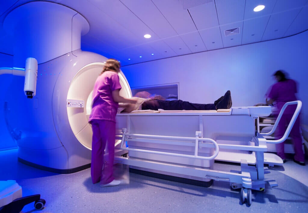
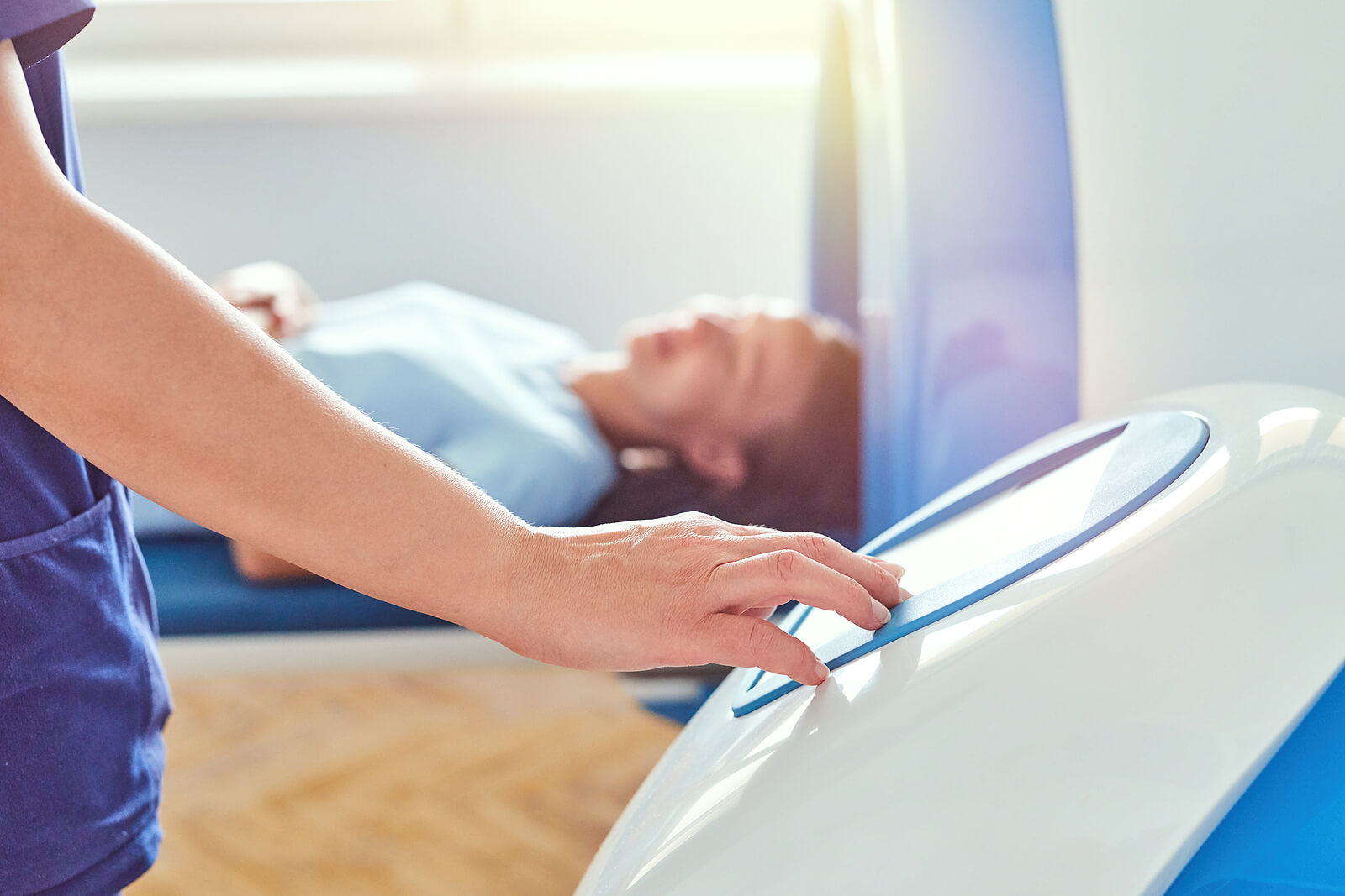
As in a CT, an MRI uses a tunnel-shaped device, in which the patient is placed on a stretcher. The contraption contains the magnets that will create the aforementioned magnetic field. Contrast fluids are sometimes necessary to strengthen the electromagnetic signals emitted from the cells of the body.
Resonance uses an electromagnetic field and a beam of radio waves. The information received is processed and displayed in the form of images on a computer.
The differences between a CT and an MRI
Now that you know how these hospital imaging techniques work in a brief way, we’re ready to explore the differences between a CT and an MRI by section. You’ll see that there are more differences than it first seems.
1. A CT uses X-rays, while MRI uses magnetic fields
A CT uses X-ray beams that are picked up by specific detectors once they pass through the patient’s body. On the other hand, resonance uses an electromagnetic field and a beam of radio waves to modify the properties of the protons in the body, thus obtaining an image when they release energy.
Although in both cases, a stretcher and a donut-shaped appliance with a central space are used, the distinction is clear. A CT makes use of X-rays, whereas an MRI doesn’t. This is vitally important in order to understand the risks and why some people benefit much more from an MRI based on the clinical picture they present.
CT scans use radiation, which can damage cells in the patient’s body.
2. The contrasts used are different
The images obtained after performing a CT scan are generally in black and white, so the structures and organs of the patient are seen in a wide grayscale. The difficulty in distinguishing nuances shows that it’s sometimes necessary to use contrasts, a series of compounds that are introduced into the body to make a structure clearly distinguishable from the rest.
There are two large groups of contrasts that can be administered before a CT scan, as indicated by the MEDSIR portal. These are the following:
- Positive contrasts: These absorb a greater amount of radiation (they’re radiopaque), so the structure of interest in which they accumulate appears whiter than the rest of the image. The most commonly used positive contrasts in this area are barium sulfate and iodine salts. Their route of entry is usually intravenous, oral, or rectal.
- Negative contrasts: These absorb less radiation (they’re radiolucent), so the structure of interest appears in the image darker in color and blacker than the rest. These are gases, such as oxygen, CO₂, or helium.
As indicated by the Doctoralia portal, resonance also benefits on many occasions from the use of contrasts, as they allow a better view of the structure to be analyzed. These are the following:
- Positive Contrasts: The most well-known positive contrast is gadolinium, a rare silver-white metallic element. Gadolinium solutions strengthen the electromagnetic signal emitted by cells, which makes the image obtained clearer and sharper.
- Negative contrasts: These consist mainly of iron. Their function is to weaken electromagnetic signals from certain areas to highlight nearby structures.
As you can see, the types of contrasts used mark another of the vital differences between a CT and an MRI. The first method uses barium sulfate, iodine salts, and gases, while the second usually uses gadolinium or iron.
3. Miscellaneous utilities
The use of each of these techniques is very varied. We’ll analyze their uses separately and then compare their differences with reference to their uses in the clinical setting.
3.1 The uses of a CT scan
The technical peculiarities of a CT allow it to be an excellent imaging method to detect certain clinical pictures. Here are some of the scenarios in which this type of test is used the most, according to the Mayo Clinic and other professional sources:
- It’s one of the fastest and most accurate tools for examining the pelvis, chest, and abdomen. This is because it provides highly detailed cross-sectional images of all types of tissue.
- It’s used in patients who’ve suffered severe contusions to detect internal injuries. A CT is very useful for emergency screening of people who’ve experienced a car accident.
- It’s helpful in discerning what’s causing abdominal pain, chest discomfort, or shortness of breath.
- CT is considered the best method to detect malignant neoplasms (cancers) of the abdomen and chest. This type of imaging technology makes it possible to quickly identify the presence of a tumor, its extent, and whether a relapse or metastasis has occurred.
- It’s used for the identification of possible vascular disorders. For example, it can detect a pulmonary embolus.
- A CT can be used to find signs compatible with the following diseases: Neuroblastoma, kidney tumors, lymphoma, cystic fibrosis, complications of appendicitis, inflammatory bowel disease, congenital malformations, and complications after pneumonia, among others.
3.2 The uses of an MRI
On the other hand, MRIs are considered to generate more detailed images than CT scans, but they’re slower, more expensive techniques and they report some logistical problems with the patient. As indicated by the Radiology.info portal, these are some of the most important uses of an MRI:
- It evaluates the state of the organs of the thorax and abdomen, including the heart, lungs, and many other structures.
- It evaluates the state of the organs located in the pelvic region, such as the bladder, uterus, ovaries, and other structures related to urination or reproduction.
- It registers abnormalities at the vascular and ganglionic levels.
- It allows the detection of malignant tumors that CT scans overlook. Prostate cancer, uterine cancer, and some types of liver cancer are virtually undetectable by CT. Also, metastasis in the brain or bone is much better seen with an MRI.
- Other pathologies detected by MRI: Cancers in the chest, abdomen, and pelvis, cirrhosis, ulcerative colitis, Crohn’s disease, heart problems, vascular inflammation, and fetal problems, among others.
3.3 The difference lies in the specificity
A CT is the first type of advanced test to be performed in all cases when cancer is suspected. In any case, if you want to detect a metastasis or a neoplasm at the pelvic or reproductive level, an MRI is usually more useful.
On the other hand, MRIs are very effective in observing the fetus inside the uterus of a pregnant woman, as they haven’t been proven to cause damage to the unborn child. Even so, it’s best for pregnant women not to undergo either of these tests until after the first trimester, as the magnetic field applied is quite strong.
A CT is used to obtain general images of certain areas, while an MRI provides greater image specificity.
4. An MRI is a more cumbersome procedure and is more expensive
After exposing these diagnostic differences between a CT and an MRI, you’re surely wondering why an MRI isn’t performed in all cases. The answer is quite simple: MRIs are considerably more expensive. A CT costs between $500 and $3,000 in the United States, while an MRI has a base price of $1,200 and goes up to $4,000.
At the same time, it’s important to emphasize that a CT scan is performed quickly, as the images are generated in a matter of seconds and the complete test rarely exceeds a time span of 20 minutes. An MRI represents a very different scenario, as it lasts from 30 to 60 minutes (or much longer) and the patient must remain completely still so that the image isn’t blurred.
In addition, it should be noted that the donut of an MRI machine is much narrower and more enclosed than the tubular structure of a CT. All this makes very sick people, the elderly, children, and those patients with anxiety or claustrophobia have a really hard time. In special cases, sedation or the oral administration of painkillers is required.
An MRI is more accurate than a CT, but it’s also considerably more expensive and technically cumbersome. People with claustrophobia or anxiety can have a very difficult time undergoing an MRI.
5. A CT scan reports a certain risk of cancer

In the first place, it’s important to emphasize that a CT will always save more lives than it can cost, but we can’t fail to mention this last point. As indicated by the United States Food and Drug Administration (FDA), the probability of developing a fatal cancer after a CT scan is 1 in 2000 patients.
The risk lies in the use of X-rays. As ionizing radiation is applied directly to the cells, there’s a very small possibility that they’ll suffer DNA damage, causing them to transform into cancer cells with unlimited growth. As an MRI doesn’t use this type of radiation, there’s no associated cancer risk.
In any case, one thing must be clear: The probability of developing cancer after a CT scan is 1 in 2000, but natural estimates are that 1 in 5 humans will naturally suffer a malignancy in their lifetime (400 for every 2000). In other words, the benefits of this test far outweigh the potential harms.
The risk of a patient developing cancer after having a CT scan is negligible, but it must be remembered anyway.
Differences between a CT and an MRI: 2 excellent imaging techniques
Here we’ve presented you with 5 differences between a CT scan and an MRI, but we don’t want you to think that one is better than the other. Although a CT is somewhat less accurate than its counterpart, it’s a cheaper, simpler, and faster diagnostic method. Therefore, it’s almost always the first option when cancer or internal injury is suspected.
An MRI provides a better image of tissues and can detect hidden neoplasms, but it’s more expensive and time-consuming to perform. Therefore, it’s carried out in more specific settings. Both techniques have their pros and cons, but your doctor will know which is the most accurate and necessary in each case.
- Definición de Tomografía Axial Computarizada, NCI. Recogido a 31 de octubre en https://www.cancer.gov/espanol/publicaciones/diccionarios/diccionario-cancer/def/tomografia-axial-computarizada
- Computed tomography (CT), NIH. Recogido a 31 de octubre en https://www.nibib.nih.gov/science-education/science-topics/computed-tomography-ct
- ¿Qué es el contraste en un TAC y para que se utiliza?, MEDSIR. Recogido a 31 de octubre en https://conviveconelcancer.com/que-es-el-contraste-en-un-tac-y-para-que-se-utiliza/
- Resonancia con contraste: ¿Qué es? Doctoralia. Recogido a 31 de octubre en https://labs.doctoralia.com.mx/blog/resonancia-magnetica-con-contraste-que-es/
- CT Scan, Mayo Clinic. Recogido a 31 de octubre en https://www.mayoclinic.org/tests-procedures/ct-scan/about/pac-20393675
- RMN de cuerpo (tórax, abdomen, pelvis), Radiology.info. Recogido a 31 de octubre en https://www.radiologyinfo.org/es/info/bodymr
- What are the radiaton risks of CT? FDA. Recogido a 31 de octubre en https://www.fda.gov/radiation-emitting-products/medical-x-ray-imaging/what-are-radiation-risks-ct