The Occipital Lobe: Characteristics and Function
The cerebral lobes are the areas of the cerebral cortex that the brain is subdivided into. We have up to 6 functional lobes, which are the occipital, frontal, parietal, temporal, insula, and limbic lobes (Huang, 2017). Today we’ll be considering the occipital lobe.
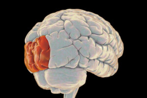
The occipital lobe, located at the back of the brain, is responsible for visually processing the information it receives. More specifically, we can say that it regulates, coordinates, and processes vision. In this article, we’ll explain how it works and what happens if it gets injured.
Occipital lobe: definition and characteristics
The occipital lobe is an area of the brain related to vision and other functions. It’s one of the smallest lobes in the brain, occupying a small area at the back of the brain. It’s located near the temporal lobe and just above the cerebellum.
There are actually two occipital lobes, one in each cerebral hemisphere. They’re almost symmetrical, and are separated by a narrow fissure.
Evolution
During its evolution, this lobe hasn’t grown in proportion to the rest of the parts of the brain, such as the frontal lobe. Neither has it become more complex over time, whereas lobes like the frontal lobe have.
As a curious fact, experts believed that Neanderthals, an evolutionary branch parallel to Homo sapiens, had a larger occipital lobe than our species.
Functioning
It’s important to know that each of the structures of our brain, such as the occipital lobe, aren’t exclusively responsible for just one function; they take care of many.
In addition, these functions are distributed throughout the brain, which means that coordination and interconnection of structures is needed for the execution of various functions.
However, what we can say is that the occipital lobe is primarily responsible for processing visual information.
Visual processing
When we see or look around us, that is, when the retinas receive visual information, it is the visual cortex that receives this information first. This cortex is included in the occipital lobe.
This cortex is divided into different areas or portions of the brain, which can be classified according to their level of visual processing.
The primary visual cortex, for example, also called V1, is the part of the occipital lobe that’s in charge of processing the “raw” visual information, as it detects the general patterns that our eyes collect.
What happens to this information? It’s then sent to other areas of the occipital lobe, areas that are responsible for processing this visual information in a more refined way. These areas, in turn, send the already analyzed information to other areas of the brain.
Ventral and dorsal pathways
The visual information has already passed through the primary visual cortex, and this area is then responsible for emitting the information via two routes: the ventral route and the dorsal route. These pathways run in parallel and, in turn, communicate with other areas of the brain:
- The ventral pathway (the what pathway): This is directed towards the frontal area of the brain, and is in charge of processing the information analyzed by V1. It processes the characteristics of the elements that we see.
- Dorsal pathway (the how pathway): This also goes to the frontal area, and processes information about the location and movement of the things we see.
Vision-related functions
Beyond visual processing, we can talk about certain more specific functions, related to vision, in which the occipital lobe intervenes:
- General visual mapping, which helps special reasoning and enhances visual memory.
- Determining the properties of the colors of the objects we see.
- Evaluating the distance, size and depth in our field of vision.
- Identifying visual stimuli (familiar faces and objects).
Epilepsy in the occipital lobe

The occipital lobe has been associated with a type of epilepsy, the so-called occipital epilepsy. Experts believe it could be at least partly to blame for it. It seems that in cases of occipital epilepsy there’s a certain genetic or biological propensity to suffer it.
First definitions and classification
The first definition of occipital epilepsy was made by Gastaut in 1982, when this doctor studied a considerable number of patients before making his findings.
In this type of condition, he found that good seizure control could be found, as well as shared EEG characteristics in all cases.
Later, in 1989, Panayiotopoulos described more cases that presented a greater breadth in symptomatology than what Gastaut had observed.
Through these two contributions from researchers, the International League Against Epilepsy in 1999 began to classify this type of epilepsy in a separate category.
Clinical features of occipital epilepsy
In a review article on occipital epilepsy, by Palacios et al. (2017), with information taken from Caraballo et al. (1998), we find the clinical characteristics (or symptoms) of occipital epilepsy:
- Ictal vomiting
- Oculocephalic tonic deviation
- Migraine episodes
- Visual crisis, secondarily generalized and partial
- Anartria and hemifacial motor crises
- Prolonged crises in wakefulness and during sleep
- Epileptic seizures that can cause neurological damage
Disorders associated with the occipital lobe (lesions)
An injury to the occipital lobe can cause several symptoms or disorders. Among them, we find instances of visual hallucinations and illusions. Hallucinations can be caused by lesions in the occipital region, but also by temporal lobe seizures.
An injured occipital lobe can also cause symptoms such as confusion, difficulty in identifying familiar shapes, colors or faces, deficits in speech processes or mathematical ability, and the appearance of involuntary movements in the eyes, among others.
Other more specific lesions, in this case, in the parietal-temporal-occipital association area (POT or PTO area), can cause speech blindness (or dyslexia) with writing problems.
Alterations in the perceptual-visual system
Lesions in this lobe can also cause subtle or more obvious changes in our visual-perceptual system. This includes visual field defects and scotomas.
A scotoma is a zone of partial, temporary or permanent blindness. In healthy people, it’s the ocular blind spot, and in people with an injury to the retina, the optimal nerve or the occipital lobe, the visual damage can be more extensive.
As we’ve seen, although it’s a small lobe, the occipital lobe has a somewhat complex function, and is responsible for regulating functions related to visual processing.
It allows us to process the objects we see, differentiate colors and movements or identify the shapes and figures of things, among others. In addition, albeit to a lesser extent, it’s also linked to other cognitive processes such as speech, memory, mathematics, logic, or writing.
Injury in this area causes various symptoms (depending on the specific area injured), such as hallucinations or difficulties in identifying faces, colors or movements.
The cerebral lobes are the areas of the cerebral cortex that the brain is subdivided into. We have up to 6 functional lobes, which are the occipital, frontal, parietal, temporal, insula, and limbic lobes (Huang, 2017). Today we’ll be considering the occipital lobe.
The occipital lobe, located at the back of the brain, is responsible for visually processing the information it receives. More specifically, we can say that it regulates, coordinates, and processes vision. In this article, we’ll explain how it works and what happens if it gets injured.
Occipital lobe: definition and characteristics
The occipital lobe is an area of the brain related to vision and other functions. It’s one of the smallest lobes in the brain, occupying a small area at the back of the brain. It’s located near the temporal lobe and just above the cerebellum.
There are actually two occipital lobes, one in each cerebral hemisphere. They’re almost symmetrical, and are separated by a narrow fissure.
Evolution
During its evolution, this lobe hasn’t grown in proportion to the rest of the parts of the brain, such as the frontal lobe. Neither has it become more complex over time, whereas lobes like the frontal lobe have.
As a curious fact, experts believed that Neanderthals, an evolutionary branch parallel to Homo sapiens, had a larger occipital lobe than our species.
Functioning
It’s important to know that each of the structures of our brain, such as the occipital lobe, aren’t exclusively responsible for just one function; they take care of many.
In addition, these functions are distributed throughout the brain, which means that coordination and interconnection of structures is needed for the execution of various functions.
However, what we can say is that the occipital lobe is primarily responsible for processing visual information.
Visual processing
When we see or look around us, that is, when the retinas receive visual information, it is the visual cortex that receives this information first. This cortex is included in the occipital lobe.
This cortex is divided into different areas or portions of the brain, which can be classified according to their level of visual processing.
The primary visual cortex, for example, also called V1, is the part of the occipital lobe that’s in charge of processing the “raw” visual information, as it detects the general patterns that our eyes collect.
What happens to this information? It’s then sent to other areas of the occipital lobe, areas that are responsible for processing this visual information in a more refined way. These areas, in turn, send the already analyzed information to other areas of the brain.
Ventral and dorsal pathways
The visual information has already passed through the primary visual cortex, and this area is then responsible for emitting the information via two routes: the ventral route and the dorsal route. These pathways run in parallel and, in turn, communicate with other areas of the brain:
- The ventral pathway (the what pathway): This is directed towards the frontal area of the brain, and is in charge of processing the information analyzed by V1. It processes the characteristics of the elements that we see.
- Dorsal pathway (the how pathway): This also goes to the frontal area, and processes information about the location and movement of the things we see.
Vision-related functions
Beyond visual processing, we can talk about certain more specific functions, related to vision, in which the occipital lobe intervenes:
- General visual mapping, which helps special reasoning and enhances visual memory.
- Determining the properties of the colors of the objects we see.
- Evaluating the distance, size and depth in our field of vision.
- Identifying visual stimuli (familiar faces and objects).
Epilepsy in the occipital lobe
The occipital lobe has been associated with a type of epilepsy, the so-called occipital epilepsy. Experts believe it could be at least partly to blame for it. It seems that in cases of occipital epilepsy there’s a certain genetic or biological propensity to suffer it.
First definitions and classification
The first definition of occipital epilepsy was made by Gastaut in 1982, when this doctor studied a considerable number of patients before making his findings.
In this type of condition, he found that good seizure control could be found, as well as shared EEG characteristics in all cases.
Later, in 1989, Panayiotopoulos described more cases that presented a greater breadth in symptomatology than what Gastaut had observed.
Through these two contributions from researchers, the International League Against Epilepsy in 1999 began to classify this type of epilepsy in a separate category.
Clinical features of occipital epilepsy
In a review article on occipital epilepsy, by Palacios et al. (2017), with information taken from Caraballo et al. (1998), we find the clinical characteristics (or symptoms) of occipital epilepsy:
- Ictal vomiting
- Oculocephalic tonic deviation
- Migraine episodes
- Visual crisis, secondarily generalized and partial
- Anartria and hemifacial motor crises
- Prolonged crises in wakefulness and during sleep
- Epileptic seizures that can cause neurological damage
Disorders associated with the occipital lobe (lesions)
An injury to the occipital lobe can cause several symptoms or disorders. Among them, we find instances of visual hallucinations and illusions. Hallucinations can be caused by lesions in the occipital region, but also by temporal lobe seizures.
An injured occipital lobe can also cause symptoms such as confusion, difficulty in identifying familiar shapes, colors or faces, deficits in speech processes or mathematical ability, and the appearance of involuntary movements in the eyes, among others.
Other more specific lesions, in this case, in the parietal-temporal-occipital association area (POT or PTO area), can cause speech blindness (or dyslexia) with writing problems.
Alterations in the perceptual-visual system
Lesions in this lobe can also cause subtle or more obvious changes in our visual-perceptual system. This includes visual field defects and scotomas.
A scotoma is a zone of partial, temporary or permanent blindness. In healthy people, it’s the ocular blind spot, and in people with an injury to the retina, the optimal nerve or the occipital lobe, the visual damage can be more extensive.
As we’ve seen, although it’s a small lobe, the occipital lobe has a somewhat complex function, and is responsible for regulating functions related to visual processing.
It allows us to process the objects we see, differentiate colors and movements or identify the shapes and figures of things, among others. In addition, albeit to a lesser extent, it’s also linked to other cognitive processes such as speech, memory, mathematics, logic, or writing.
Injury in this area causes various symptoms (depending on the specific area injured), such as hallucinations or difficulties in identifying faces, colors or movements.
- Caraballo, R., Cersosimo, R., Medina, C., Tenembaum, S. y Fejerman, N. (1998). Epilepsias occipitales idiopáticas. Arch Arg Pediatr, 96: 169-176.
- Carlson, Neil R. (2007). Psychology : the science of behaviour. New Jersey, USA: Pearson Education.
- Carlson, N.R. (1999). Fisiología de la conducta. Barcelona: Ariel Psicología.
- Carpenter, M.B. (1994). Neuroanatomía. Fundamentos. Buenos Aires: Editorial Panamericana.
- Destina Yalçin, A.; Kaymaz, A.; Forta, H. (2000). Reflex occipital lobe epilepsy. Seizure.
- Huang J. (2017). Revisión sobre la función cerebral. Manual MSD. Versión para profesionales.
- Palacios, E., Bello, L., Maldonado, D. y Martínez, F. (2017). Epilepsia occipital. Repertorio de Medicina y Cirugía, 26(1): 3-8.
Este texto se ofrece únicamente con propósitos informativos y no reemplaza la consulta con un profesional. Ante dudas, consulta a tu especialista.


