Meninges: Characteristics and Functions
The central nervous system (CNS) is made up of the brain and spinal cord, which are very delicate structures. The importance of these bodies makes it necessary to have different protection measures. At this point, we have the meninges, a set of layers that protect the CNS from physical and chemical damage.
The bones of the skull and spinal column are the main means of protection for the structures that make up the CNS. They provide a hard and resistant cover capable of withstanding blows of a certain magnitude. However, these structures are in constant motion, and so they need a damping system.
The meninges have various structures such as venous plexuses and sensory innervation. However, the diseases that affect them are very dangerous and can cause death. We’re now going to talk about the main characteristics and the most relevant functions of these layers.
What are the meninges?
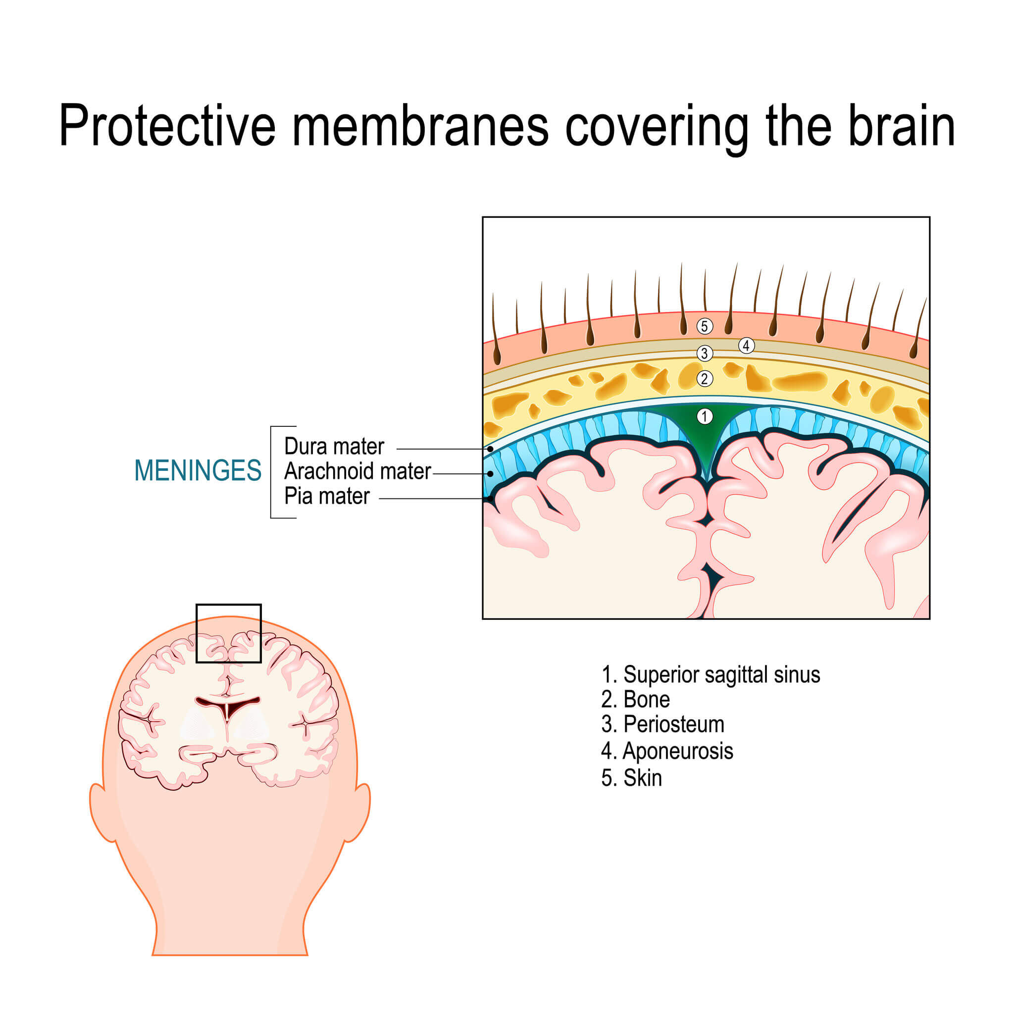
The meninges are one of the tissues that are responsible for providing protection to the brain and spinal cord. They’re made up of 3 connective tissue membranes, located one on top of the other, surrounding all the brain and spinal structures.
These structures are essential organs for life, and are responsible for generating and transmitting orders for the proper functioning of the body. Both organs can be considered as the command center and the communication channel respectively, and, because of this, damage to them can be catastrophic.
The three layers that surround the CNS are the dura mater, arachnoid mater, and pia mater. In addition, these structures have anatomical spaces between them that have physiological functions and are of great importance in the medical field.
Dura mater
When it comes to accessing the central nervous system from the outside in, the first layer of the meninges that can be seen is the dura mater. This is the hardest and most resistant layer of all, as it’s the one that’s in intimate contact with the protective bone.
The layer in question is made up of multiple lamellae of collagen and elastin fibers, which are located in sets and can measure up to 5 microns according to observations in an electron microscope. For its part, this layer also helps the division of various structures in the brain as it forms the following partitions:
- Sickle of the brain: This divides the brain into two hemispheres.
- Cerebellar tentorium: This separates the occipital lobes from the rest of the brain and delimits the trigeminal nerve.
- Sickle cerebellum: This divides the cerebellum into two different hemispheres.
- Pituitary tentorium: This is a septum that surrounds the sella turcica and protects the organ called the pituitary.
On the spinal cord, there’s an anatomical space between the dura mater and the spinal column, called the epidural space. It contains loose connective tissue, adipose tissue, and the internal vertebral venous plexus. The anatomical importance of this space is that it is the ideal place to inject local anesthetics in the lumbar and sacral region to facilitate various medical processes.
Arachnoid mater
The arachnoid mater is the second layer of the meninges, and it’s in contact with both the dura mater and the pia mater. It’s the thinnest and weakest layer of all, and so it’s prone to breakage. In addition, another of its main characteristics is that it isn’t vascularized.
This layer is made up of connective tissue and serous portions that allow it to perform its function adequately. The arachnoid mater is composed of two different layers, the arachnoid barrier, and the arachnoid trabeculae. The first one is in contact with the dura mater; it‘s the most resistant part and prevents the entry of ions and other molecules.
The arachnoid trabeculae are evaginations that come into contact with the pia mater and facilitate its communication with the arachnoid mater. All these trabeculae form a kind of network that crosses the subarachnoid space, which contains anchoring fibers and microfibers.
Between both layers is a region called the subarachnoid space, through which the cerebrospinal fluid circulates, a substance that has a precise and sterile chemical composition. Within this space, there are also various cavities where the liquid in question accumulates and is distributed.
Pia mater
Last of all, we have the deepest layer of the meninges, called the pia mater, which is in intimate contact with the brain and medullary surface. It’s composed of serous tissue, and so it’s very fine and has the consistency of a flexible mesh.
Some studies show that this membrane is very permeable compared to the previous two, thanks to the natural perforations it has, especially in the lumbar region.
The pia mater contains structures called choroidal folds, which join the choroid plexus, which is the area where cerebrospinal fluid is secreted, which is essential for shock absorption. This membrane also supports the blood vessels that enter the brain to nourish it.
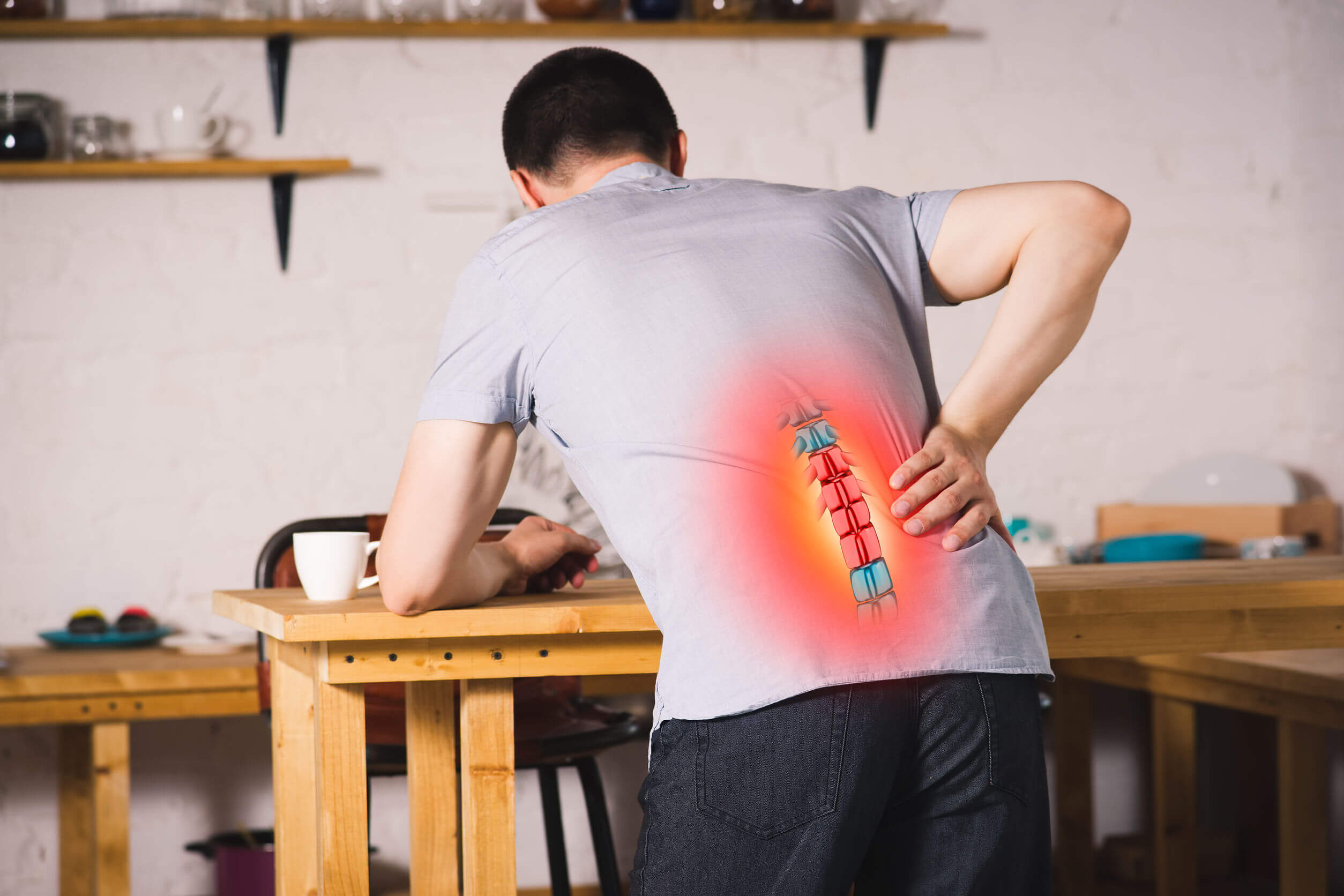
On the other hand, this membrane is in contact with nociceptors, which are the receptors responsible for perceiving pain. This characteristic is useful when a person has herniated discs, as it will be an indication that something isn’t working properly.
Functions of the meninges
The main function of these three structures is to protect the central nervous system, forming a physical protection mechanism against trauma and microorganisms.
All these membranes form the blood-brain barrier, and so they have a very selective permeability and prevent the passage of substances that may be harmful. In this way, there are a rather limited number of molecules that are capable of passing through them. In addition to this, microorganisms can’t penetrate this barrier either, and so many infections are prevented.
Lastly, all the meninges provide a perfect cushioning system for the brain and spinal cord. This occurs thanks to the cerebrospinal fluid contained in the subarachnoid space, which will minimize the movement of the structures during trauma or some physical activity.
In general terms, the meninges form a kind of support around the CNS, preventing it from deforming.
The meninges: the last layer of protection of the CNS
The membranes in question can be defined as the last measure of protection for the CNS. This is because they are the last tissues that you’ll find before coming into contact with the nervous system. Despite being very thin layers, they’re essential for life.
The meninges form one of the most difficult to penetrate barriers in the whole of the body, thus protecting the CNS from both foreign molecules and harmful pathogens. However, these membranes can be affected by pathologies such as meningitis, which constitute a true medical emergency.
The central nervous system (CNS) is made up of the brain and spinal cord, which are very delicate structures. The importance of these bodies makes it necessary to have different protection measures. At this point, we have the meninges, a set of layers that protect the CNS from physical and chemical damage.
The bones of the skull and spinal column are the main means of protection for the structures that make up the CNS. They provide a hard and resistant cover capable of withstanding blows of a certain magnitude. However, these structures are in constant motion, and so they need a damping system.
The meninges have various structures such as venous plexuses and sensory innervation. However, the diseases that affect them are very dangerous and can cause death. We’re now going to talk about the main characteristics and the most relevant functions of these layers.
What are the meninges?
The meninges are one of the tissues that are responsible for providing protection to the brain and spinal cord. They’re made up of 3 connective tissue membranes, located one on top of the other, surrounding all the brain and spinal structures.
These structures are essential organs for life, and are responsible for generating and transmitting orders for the proper functioning of the body. Both organs can be considered as the command center and the communication channel respectively, and, because of this, damage to them can be catastrophic.
The three layers that surround the CNS are the dura mater, arachnoid mater, and pia mater. In addition, these structures have anatomical spaces between them that have physiological functions and are of great importance in the medical field.
Dura mater
When it comes to accessing the central nervous system from the outside in, the first layer of the meninges that can be seen is the dura mater. This is the hardest and most resistant layer of all, as it’s the one that’s in intimate contact with the protective bone.
The layer in question is made up of multiple lamellae of collagen and elastin fibers, which are located in sets and can measure up to 5 microns according to observations in an electron microscope. For its part, this layer also helps the division of various structures in the brain as it forms the following partitions:
- Sickle of the brain: This divides the brain into two hemispheres.
- Cerebellar tentorium: This separates the occipital lobes from the rest of the brain and delimits the trigeminal nerve.
- Sickle cerebellum: This divides the cerebellum into two different hemispheres.
- Pituitary tentorium: This is a septum that surrounds the sella turcica and protects the organ called the pituitary.
On the spinal cord, there’s an anatomical space between the dura mater and the spinal column, called the epidural space. It contains loose connective tissue, adipose tissue, and the internal vertebral venous plexus. The anatomical importance of this space is that it is the ideal place to inject local anesthetics in the lumbar and sacral region to facilitate various medical processes.
Arachnoid mater
The arachnoid mater is the second layer of the meninges, and it’s in contact with both the dura mater and the pia mater. It’s the thinnest and weakest layer of all, and so it’s prone to breakage. In addition, another of its main characteristics is that it isn’t vascularized.
This layer is made up of connective tissue and serous portions that allow it to perform its function adequately. The arachnoid mater is composed of two different layers, the arachnoid barrier, and the arachnoid trabeculae. The first one is in contact with the dura mater; it‘s the most resistant part and prevents the entry of ions and other molecules.
The arachnoid trabeculae are evaginations that come into contact with the pia mater and facilitate its communication with the arachnoid mater. All these trabeculae form a kind of network that crosses the subarachnoid space, which contains anchoring fibers and microfibers.
Between both layers is a region called the subarachnoid space, through which the cerebrospinal fluid circulates, a substance that has a precise and sterile chemical composition. Within this space, there are also various cavities where the liquid in question accumulates and is distributed.
Pia mater
Last of all, we have the deepest layer of the meninges, called the pia mater, which is in intimate contact with the brain and medullary surface. It’s composed of serous tissue, and so it’s very fine and has the consistency of a flexible mesh.
Some studies show that this membrane is very permeable compared to the previous two, thanks to the natural perforations it has, especially in the lumbar region.
The pia mater contains structures called choroidal folds, which join the choroid plexus, which is the area where cerebrospinal fluid is secreted, which is essential for shock absorption. This membrane also supports the blood vessels that enter the brain to nourish it.
On the other hand, this membrane is in contact with nociceptors, which are the receptors responsible for perceiving pain. This characteristic is useful when a person has herniated discs, as it will be an indication that something isn’t working properly.
Functions of the meninges
The main function of these three structures is to protect the central nervous system, forming a physical protection mechanism against trauma and microorganisms.
All these membranes form the blood-brain barrier, and so they have a very selective permeability and prevent the passage of substances that may be harmful. In this way, there are a rather limited number of molecules that are capable of passing through them. In addition to this, microorganisms can’t penetrate this barrier either, and so many infections are prevented.
Lastly, all the meninges provide a perfect cushioning system for the brain and spinal cord. This occurs thanks to the cerebrospinal fluid contained in the subarachnoid space, which will minimize the movement of the structures during trauma or some physical activity.
In general terms, the meninges form a kind of support around the CNS, preventing it from deforming.
The meninges: the last layer of protection of the CNS
The membranes in question can be defined as the last measure of protection for the CNS. This is because they are the last tissues that you’ll find before coming into contact with the nervous system. Despite being very thin layers, they’re essential for life.
The meninges form one of the most difficult to penetrate barriers in the whole of the body, thus protecting the CNS from both foreign molecules and harmful pathogens. However, these membranes can be affected by pathologies such as meningitis, which constitute a true medical emergency.
- Reina M, López A, Dittman M, De Andrés J. Análisis estructural del espesor de la duramadre humana mediante microscopía electrónica de barrido. Rev Esp Anestesiol Reanim. 1996; 43(4), 135-137.
- Reina M, Pulido P, López A. El saco dural humano. Morfología de la dura-aracnoides espinal. Origen del espacio subduralespinal. Rev Argent Anest. 2007; 65: 167-84.
- Reina M, García A, De Andres J. Descripción anatómica de la presencia de perforaciones naturales en la piamadre humana en la médula lumbar. Rev. Esp. Anestesiol. Reánim. 1998; 45: 4-7.
- Coles JA, Stewart-Hutchinson PJ, Myburgh E, Brewer JM. The mouse cortical meninges are the site of immune responses to many different pathogens, and are accessible to intravital imaging. Methods. 2017;127:53-61.
- Sze G. Diseases of the intracranial meninges: MR imaging features. AJR Am J Roentgenol. 1993;160(4):727-33.
- Meltzer CC, Fukui MB, Kanal E, Smirniotopoulos JG. MR imaging of the meninges. Part I. Normal anatomic features and nonneoplastic disease. Radiology. 1996;201(2):297-308.
Este texto se ofrece únicamente con propósitos informativos y no reemplaza la consulta con un profesional. Ante dudas, consulta a tu especialista.


