Uterine Fibroids: Symptoms, Causes and Treatment
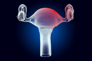
A myoma is defined as ‘the benign and abnormal growth of muscle cells of a specific organ’. Uterine fibroids are the most frequent tumors in the area of gynecology, and can affect millions of women around the world. Do you want to know what its symptoms, causes, and treatment are? Keep reading!
Multiple studies affirm that these tumors are present in up to 40% of women under 40 years of age worldwide. These tumors appear in the muscular layer of the uterus, and are largely composed of multiple layers of collagen along with other substances such as fibronectin and proteoglycans.
Uterine fibroids can appear in any woman, although they’re more common in African-Americans than Caucasians. Their characteristics may vary from person to person. In this sense, they can be unique, multiple, so small that they’re undetectable or so large that they produce compression.
Symptoms

Although neoplasms (tumors) are associated with severe symptoms and pathologies, research shows that most uterine fibroids are asymptomatic. In fact, it’s estimated that only between 20 and 50% of women have some type of symptom. The appearance of these symptoms is related to the size, location and number of tumors.
The most common symptom is the appearance of abnormal uterine bleeding. A very common form of bleeding is metrorrhagia, that is, vaginal bleeding that doesn’t coincide with the days of menstruation. Very heavy menstrual bleeding, known as hypermenorrhea, can also occur.
The presence of multiple uterine fibroids and their increase in size is related to the appearance of a very diverse clinical picture. In this sense, women may show the following symptoms:
- The need to urinate more often
- Pain or pressure in the pelvis
- Infertility
- Pain during sexual intercourse or dyspareunia
- Constipation
Causes of uterine fibroids
Unfortunately, a specific cause that explains the appearance of uterine fibroids hasn’t yet been established. We currently know that they appear as a result of a monoclonal alteration, that is, that they’re due to the growth and multiplication of a single muscle cell.
The appearance of uterine fibroids has been related to the action of circulating estrogens and progesterone on the uterine muscle due to the age at which they appear. These alterations are very rare during puberty, while they increase in frequency between the ages of 40 and 50.
Many different studies relate the appearance of uterine fibroids with alterations in different chromosomes, among which we find 1, 3, 7, 12, and 14. In fact, it has been shown that variations in these chromosomes have several origins, from translocation of its portions to the deletion of some gene (modifications in the DNA).
Risk factors
On the other hand, there are many risk factors that increase the likelihood of fibroids appearing in the uterus. Many of them are related to increased levels of estrogen in the blood and the following stand out:
- Being between 40 and 50 years of age
- Being of African descent
- Using oral contraceptives
- The appearance of obesity and having a sedentary lifestyle
- Carrying out hormone replacement therapy
- Having a family history of uterine fibroids
- Smoking frequently
Some of these risk factors are related to the genetics of women, so they can’t be modified. However, most of them depend on a person’s lifestyle, so they can be modified. In this sense, it’s possible to reduce the probability of the appearance of uterine fibroids.
Diagnosis
The diagnosis of uterine fibroids can be very difficult, since most of them are only a few centimeters long and are asymptomatic. The doctor may suspect the presence of these changes during a routine pelvic examination, especially if they have a significant diameter.
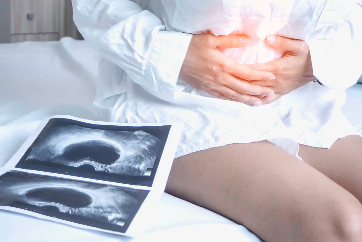
Given the suspicion of a uterine myoma, the specialist will explain the different gynecological tests that are available, which can give a definitive diagnosis. Pelvic or transvaginal ultrasound is one of the most useful imaging tests in this type of pathology. It uses waves to obtain an image of the uterus and makes it possible to measure fibroids.
In some cases, the ultrasound may not show the tumors clearly, and so it’s necessary to perform other types of studies. Among the other imaging tests that can be used, the following stand out:
- Magnetic resonance
- Hysteroecography
- Hysterosalpingography
- Hysteroscopy
Blood tests are useful when establishing differential diagnoses and ruling out other pathologies that cause menstrual alterations. In this regard, it’s possible to indicate a complete blood count and analysis of the levels of various substances such as thyroid hormones.
Treatment of uterine fibroids
The treatment of uterine fibroids will depend on the characteristics of the tumors and the presence or absence of symptoms. In this sense, asymptomatic fibroids don’t merit any type of treatment and should only be evaluated from time to time.
On the other hand, in the presence of annoying symptoms, there can be two approaches, one conservative and the other surgical. Both approaches have very good results and help to improve the patient’s quality of life.
Conservative treatment
This therapeutic option is intended for women with moderate-sized fibroids whose symptoms aren’t severe. Treatment will be aimed at reducing estrogen levels in order to prevent tumor growth and reduce the intensity of symptoms.
In this sense, hormone-releasing intrauterine devices can be placed, oral contraceptives administered, or hormonal therapy injections are given. In addition, drugs can be used to limit blood flow to the fibroid, as well as to use compounds to relieve pain and treat other specific symptoms.
Surgical treatment
Surgical removal of uterine fibroids is indicated in those patients with neoplasms of considerable size or with severe symptoms. The procedure to be carried out will depend on the age of the patient, her physical condition and the number and size of the tumors.
Generally speaking, the procedure will seek to remove the entire fibroid while preserving as much of the uterus as possible.
The techniques that can be used are very varied, they range from arterial embolization to reduce blood flow to the fibroid and cause tissue death (necrosis), to performing a hysterectomy in the most severe cases.
A common but benign pathology
Uterine fibroids represent the most common cause of pelvic tumors in women of childbearing age. They may appear as a result of the action of estrogens on the cells of the muscular layer of the uterus. In addition, their appearance is influenced by multiple risk factors that depend on a person’s lifestyle.
Some of these tumors can cause very uncomfortable symptoms in those who suffer from them, and there’s even the possibility that they may affect fertility and compress other organs. All of these tumors are benign and the chance of them turning into cancer is very low. However, it’s important to visit your doctor if you have symptoms that concern you.
A myoma is defined as ‘the benign and abnormal growth of muscle cells of a specific organ’. Uterine fibroids are the most frequent tumors in the area of gynecology, and can affect millions of women around the world. Do you want to know what its symptoms, causes, and treatment are? Keep reading!
Multiple studies affirm that these tumors are present in up to 40% of women under 40 years of age worldwide. These tumors appear in the muscular layer of the uterus, and are largely composed of multiple layers of collagen along with other substances such as fibronectin and proteoglycans.
Uterine fibroids can appear in any woman, although they’re more common in African-Americans than Caucasians. Their characteristics may vary from person to person. In this sense, they can be unique, multiple, so small that they’re undetectable or so large that they produce compression.
Symptoms
Although neoplasms (tumors) are associated with severe symptoms and pathologies, research shows that most uterine fibroids are asymptomatic. In fact, it’s estimated that only between 20 and 50% of women have some type of symptom. The appearance of these symptoms is related to the size, location and number of tumors.
The most common symptom is the appearance of abnormal uterine bleeding. A very common form of bleeding is metrorrhagia, that is, vaginal bleeding that doesn’t coincide with the days of menstruation. Very heavy menstrual bleeding, known as hypermenorrhea, can also occur.
The presence of multiple uterine fibroids and their increase in size is related to the appearance of a very diverse clinical picture. In this sense, women may show the following symptoms:
- The need to urinate more often
- Pain or pressure in the pelvis
- Infertility
- Pain during sexual intercourse or dyspareunia
- Constipation
Causes of uterine fibroids
Unfortunately, a specific cause that explains the appearance of uterine fibroids hasn’t yet been established. We currently know that they appear as a result of a monoclonal alteration, that is, that they’re due to the growth and multiplication of a single muscle cell.
The appearance of uterine fibroids has been related to the action of circulating estrogens and progesterone on the uterine muscle due to the age at which they appear. These alterations are very rare during puberty, while they increase in frequency between the ages of 40 and 50.
Many different studies relate the appearance of uterine fibroids with alterations in different chromosomes, among which we find 1, 3, 7, 12, and 14. In fact, it has been shown that variations in these chromosomes have several origins, from translocation of its portions to the deletion of some gene (modifications in the DNA).
Risk factors
On the other hand, there are many risk factors that increase the likelihood of fibroids appearing in the uterus. Many of them are related to increased levels of estrogen in the blood and the following stand out:
- Being between 40 and 50 years of age
- Being of African descent
- Using oral contraceptives
- The appearance of obesity and having a sedentary lifestyle
- Carrying out hormone replacement therapy
- Having a family history of uterine fibroids
- Smoking frequently
Some of these risk factors are related to the genetics of women, so they can’t be modified. However, most of them depend on a person’s lifestyle, so they can be modified. In this sense, it’s possible to reduce the probability of the appearance of uterine fibroids.
Diagnosis
The diagnosis of uterine fibroids can be very difficult, since most of them are only a few centimeters long and are asymptomatic. The doctor may suspect the presence of these changes during a routine pelvic examination, especially if they have a significant diameter.
Given the suspicion of a uterine myoma, the specialist will explain the different gynecological tests that are available, which can give a definitive diagnosis. Pelvic or transvaginal ultrasound is one of the most useful imaging tests in this type of pathology. It uses waves to obtain an image of the uterus and makes it possible to measure fibroids.
In some cases, the ultrasound may not show the tumors clearly, and so it’s necessary to perform other types of studies. Among the other imaging tests that can be used, the following stand out:
- Magnetic resonance
- Hysteroecography
- Hysterosalpingography
- Hysteroscopy
Blood tests are useful when establishing differential diagnoses and ruling out other pathologies that cause menstrual alterations. In this regard, it’s possible to indicate a complete blood count and analysis of the levels of various substances such as thyroid hormones.
Treatment of uterine fibroids
The treatment of uterine fibroids will depend on the characteristics of the tumors and the presence or absence of symptoms. In this sense, asymptomatic fibroids don’t merit any type of treatment and should only be evaluated from time to time.
On the other hand, in the presence of annoying symptoms, there can be two approaches, one conservative and the other surgical. Both approaches have very good results and help to improve the patient’s quality of life.
Conservative treatment
This therapeutic option is intended for women with moderate-sized fibroids whose symptoms aren’t severe. Treatment will be aimed at reducing estrogen levels in order to prevent tumor growth and reduce the intensity of symptoms.
In this sense, hormone-releasing intrauterine devices can be placed, oral contraceptives administered, or hormonal therapy injections are given. In addition, drugs can be used to limit blood flow to the fibroid, as well as to use compounds to relieve pain and treat other specific symptoms.
Surgical treatment
Surgical removal of uterine fibroids is indicated in those patients with neoplasms of considerable size or with severe symptoms. The procedure to be carried out will depend on the age of the patient, her physical condition and the number and size of the tumors.
Generally speaking, the procedure will seek to remove the entire fibroid while preserving as much of the uterus as possible.
The techniques that can be used are very varied, they range from arterial embolization to reduce blood flow to the fibroid and cause tissue death (necrosis), to performing a hysterectomy in the most severe cases.
A common but benign pathology
Uterine fibroids represent the most common cause of pelvic tumors in women of childbearing age. They may appear as a result of the action of estrogens on the cells of the muscular layer of the uterus. In addition, their appearance is influenced by multiple risk factors that depend on a person’s lifestyle.
Some of these tumors can cause very uncomfortable symptoms in those who suffer from them, and there’s even the possibility that they may affect fertility and compress other organs. All of these tumors are benign and the chance of them turning into cancer is very low. However, it’s important to visit your doctor if you have symptoms that concern you.
- Ortiz Ruiz M, Matute Labrador A, Martínez Consuegra N. Miomatosis uterina. An Med (Mex) 2009; 54 (4): 222-233.
- Fábregues F, Peñarrubia J. Mioma uterino. Manifestaciones clínicas y posibilidades actuales de tratamiento conservador. Medicina Integral. 2002; 40 (5): 190-195.
- Sepúlveda J, Alarcón M. Manejo médico de la miomatosis uterina. Revisión de la literatura. Rev Chil Obstet Ginecol. 2016; 81(1): 48-55.
- Parker WH. Etiology, symptomatology, and diagnosis of uterine myomas. Fertil Steril. 2007;87(4):725-36.
- Fascilla FD, Cramarossa P, Cannone R, Olivieri C, Vimercati A, Exacoustos C. Ultrasound diagnosis of uterine myomas. Minerva Ginecol. 2016;68(3):297-312.
- Sparic R, Mirkovic L, Malvasi A, Tinelli A. Epidemiology of Uterine Myomas: A Review. Int J Fertil Steril. 2016;9(4):424-35.
Este texto se ofrece únicamente con propósitos informativos y no reemplaza la consulta con un profesional. Ante dudas, consulta a tu especialista.


