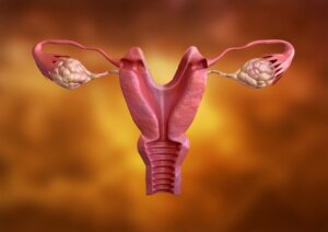
Types of Uterus and Uterine Malformations

In women, there are different types of uterus although its function is always the same. In general, these derive from “uterine malformations” produced in the embryonic stage during the development of the paramesonephric ducts or “Müllerian ducts”.
Uterine malformations are usually asymptomatic and aren’t diagnosed until a routine transvaginal ultrasound is performed or in the presence of fertility problems. These congenital anomalies occur in up to 3 to 5% of the general population.
Origin of the types of uterus
Uterine malformations develop during the embryonic stage. At this time the uterus develops as two separate halves that later merge, and when this occurs differently, the anomaly occurs.
Although uterine malformations are rare in women with infertility, it increases to 8%, and is the cause of up to 18% of repeat miscarriages as well as 25% of late miscarriages.
Classification of types of uterus

According to the European Society of Human Reproduction and Embryology (ESHRE) and the European Society of Gynecological Endoscopy (ESGE), in 2013 the following classification of uterine malformations was proposed:
- Class 0 (normal): The fundal contour is straight or curved with a midline fundal indentation that doesn’t exceed 50% of the uterine wall thickness.
- Class I (dysmorphic): On the outside it appears normal, but internally the walls of the uterus are very thick, so it’s usually very narrow or T-shaped.
- Class II (septate): It’s also normal on the outside, but on the inside it has a partition or septum that partially or totally divides the uterus into two cavities.
- Class III (bicornuate or bicorporal): It has a fold towards the interior of the uterine cavity that partially or totally divides it.
- Class IV (hemiuterus): Only one side or uterine cavity has developed, which is normal, but the other is incomplete or absent.
- Class V (aplastic or dysplastic): There is complete or unilateral absence of a fully developed uterine cavity. There are one or two rudimentary horns that don’t connect with the cavity.
- Class VI (not yet classified): Includes all rare anomalies such as subtle changes or a combination of pathologies that cannot be included in any of the previous groups.
In this case, the frequency follows an order from highest to lowest. The most frequent uterine anomaly is the dysmorphic uterus, and the least frequent is the one that cannot be classified.
There is a different classification
There’s also the classification of uterine congenital anomalies by the American Society for Reproductive Medicine revised in 2016. However, this classifies only the uterine body, unlike the aforementioned classification, which does so with the body and neck of the uterus, and vagina, allowing a diagnosis of the malformation.
Uterine malformations are usually asymptomatic
The different types of uterus usually aren’t diagnosed until a routine transvaginal ultrasound is performed or in the presence of problems conceiving.
However, in cases of hemiuterus, in which the horn doesn’t connect with the uterine cavity in the rudimentary cavity, pelvic and abdominal pain may occur. This occurs because the blood from menstruation cannot flow to the vagina and accumulates in the uterus.
The presence of uterine malformations can cause complications in pregnancy such as late or repeated miscarriages, premature births, ectopic pregnancies, third-trimester bleeding, fetal malpositions, and uterine contraction disorders during labor that make delivery difficult.
Diagnosis is with imaging studies
When in the embryonic stage there are alterations in the development of the paramesonephric ducts. There are alterations in the formation of the uterine tubes, uterovaginal canal, and in the upper part of the vagina.
To diagnose malformations, transvaginal ultrasound is used in the first instance. It’s recommended to carry this out at the end of menstruation when the upper layers of the endometrium have already been expelled.
However, in other cases, other tests such as MRI, hysteroscopy and hysterosalpingography may be necessary. Magnetic resonance imaging is the ideal study in the case of imperforate hymen, and hysterosalpingography is carried out otherwise.
Hysterosalpingography is the most widely used method to assess the status of the uterine tubes, intrauterine septa, intrauterine adhesions, submucosal fibroids, and endometrial polyps.
Should uterine malformations be treated?

The different types of uterus don’t always require surgical resolution. In patients with uterine malformations, surgical treatment is limited to those who have recurrent miscarriages with no other apparent cause.
This is also true in cases of chronic pelvic pain when it has been confirmed by laparoscopy that there is no coexistence of endometriosis. The best results are in the septate and bicorporal uterus.
When the transport of sperm and eggs is prevented, the option is in vitro fertilization.
In cases of infertility, malformations must be ruled out
Although rare in the general population, uterine malformations are more common in cases of infertility or recurrent miscarriages. Going to a gynecologist is essential if any of these disorders are suspected.
In women, there are different types of uterus although its function is always the same. In general, these derive from “uterine malformations” produced in the embryonic stage during the development of the paramesonephric ducts or “Müllerian ducts”.
Uterine malformations are usually asymptomatic and aren’t diagnosed until a routine transvaginal ultrasound is performed or in the presence of fertility problems. These congenital anomalies occur in up to 3 to 5% of the general population.
Origin of the types of uterus
Uterine malformations develop during the embryonic stage. At this time the uterus develops as two separate halves that later merge, and when this occurs differently, the anomaly occurs.
Although uterine malformations are rare in women with infertility, it increases to 8%, and is the cause of up to 18% of repeat miscarriages as well as 25% of late miscarriages.
Classification of types of uterus

According to the European Society of Human Reproduction and Embryology (ESHRE) and the European Society of Gynecological Endoscopy (ESGE), in 2013 the following classification of uterine malformations was proposed:
- Class 0 (normal): The fundal contour is straight or curved with a midline fundal indentation that doesn’t exceed 50% of the uterine wall thickness.
- Class I (dysmorphic): On the outside it appears normal, but internally the walls of the uterus are very thick, so it’s usually very narrow or T-shaped.
- Class II (septate): It’s also normal on the outside, but on the inside it has a partition or septum that partially or totally divides the uterus into two cavities.
- Class III (bicornuate or bicorporal): It has a fold towards the interior of the uterine cavity that partially or totally divides it.
- Class IV (hemiuterus): Only one side or uterine cavity has developed, which is normal, but the other is incomplete or absent.
- Class V (aplastic or dysplastic): There is complete or unilateral absence of a fully developed uterine cavity. There are one or two rudimentary horns that don’t connect with the cavity.
- Class VI (not yet classified): Includes all rare anomalies such as subtle changes or a combination of pathologies that cannot be included in any of the previous groups.
In this case, the frequency follows an order from highest to lowest. The most frequent uterine anomaly is the dysmorphic uterus, and the least frequent is the one that cannot be classified.
There is a different classification
There’s also the classification of uterine congenital anomalies by the American Society for Reproductive Medicine revised in 2016. However, this classifies only the uterine body, unlike the aforementioned classification, which does so with the body and neck of the uterus, and vagina, allowing a diagnosis of the malformation.
Uterine malformations are usually asymptomatic
The different types of uterus usually aren’t diagnosed until a routine transvaginal ultrasound is performed or in the presence of problems conceiving.
However, in cases of hemiuterus, in which the horn doesn’t connect with the uterine cavity in the rudimentary cavity, pelvic and abdominal pain may occur. This occurs because the blood from menstruation cannot flow to the vagina and accumulates in the uterus.
The presence of uterine malformations can cause complications in pregnancy such as late or repeated miscarriages, premature births, ectopic pregnancies, third-trimester bleeding, fetal malpositions, and uterine contraction disorders during labor that make delivery difficult.
Diagnosis is with imaging studies
When in the embryonic stage there are alterations in the development of the paramesonephric ducts. There are alterations in the formation of the uterine tubes, uterovaginal canal, and in the upper part of the vagina.
To diagnose malformations, transvaginal ultrasound is used in the first instance. It’s recommended to carry this out at the end of menstruation when the upper layers of the endometrium have already been expelled.
However, in other cases, other tests such as MRI, hysteroscopy and hysterosalpingography may be necessary. Magnetic resonance imaging is the ideal study in the case of imperforate hymen, and hysterosalpingography is carried out otherwise.
Hysterosalpingography is the most widely used method to assess the status of the uterine tubes, intrauterine septa, intrauterine adhesions, submucosal fibroids, and endometrial polyps.
Should uterine malformations be treated?

The different types of uterus don’t always require surgical resolution. In patients with uterine malformations, surgical treatment is limited to those who have recurrent miscarriages with no other apparent cause.
This is also true in cases of chronic pelvic pain when it has been confirmed by laparoscopy that there is no coexistence of endometriosis. The best results are in the septate and bicorporal uterus.
When the transport of sperm and eggs is prevented, the option is in vitro fertilization.
In cases of infertility, malformations must be ruled out
Although rare in the general population, uterine malformations are more common in cases of infertility or recurrent miscarriages. Going to a gynecologist is essential if any of these disorders are suspected.
- Beguería R, Checa M, Castillo M, del Amo E, Carreras R. Malformaciones Müllerianas: clasificación, diagnóstico y manejo. Ginecología y Obstetricia Clínica 2009;10(3). Disponible en https://www.researchgate.net/publication/237594936_Malformaciones_Mullerianas_clasificacion_diagnostico_y_manejo.
- Bendifallah S, Silberstein M, Levaillant J, Fernández H. Malformaciones uterovaginales y fertilidad. EMC – Ginecología-Obsetricia 2015;51(3). Disponible en https://www.sciencedirect.com/science/article/abs/pii/S1283081X15728341.
- Díaz J, Díaz J, Sierra L, Perez F. Útero bicorporeo – doble cérvix con embarazo gemelar en una sola cavidad uterina. Reporte de caso. Rev. chil. obstet. ginecol. 2022;87(5): 356-359. Disponible en: http://www.scielo.cl/scielo.php?script=sci_arttext&pid=S0717-75262022000500356&lng=es.
- Saravelos S, Cocksedge K, Li T. Prevalence and diagnosis of congenital uterine anomalies in women with reproductive failure: a critical appraisal. Human Reproductive Update 2008;14(5). Disponible en https://pubmed.ncbi.nlm.nih.gov/18539641/.
- Velandia M, Sepúlveda A. Revisión de la clasificación y diagnóstico de malformaciones mullerianas. Medicas UIS 2018;31(2). Disponible en: http://www.scielo.org.co/scielo.php?script=sci_arttext&pid=S0121-03192018000200057&lng=en.
Este texto se ofrece únicamente con propósitos informativos y no reemplaza la consulta con un profesional. Ante dudas, consulta a tu especialista.


