3D Mammography: Everything You Need to Know
Imaging studies allow experts to explore the interior of the human body in search of structural alterations or abnormal proliferations. Currently, X-rays, ultrasound and CT scans are the most widely used imaging methods. 3D mammography is one of the most recent techniques. Here we’ll tell you everything you need to know.
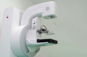
3D mammography, also called breast tomosynthesis, is a type of x-ray that combines multiple x-ray slices to provide a detailed, three-dimensional image of the breast.
Why is it performed?

In general, 3D mammography is used as an early detection test for breast cancer, especially in people with risk factors who don’t have signs of the disease.
Studies claim that 3D mammography offers up to 90% sensitivity for breast cancer and is capable of detecting up to 40% more cancer cases than standard digital mammography.
The images offered by this test are processed in a similar way to computed tomography, which provides greater detail in the detection of lesions or tumor processes. Likewise, this test reduces the need for other additional imaging studies thanks to its ability to take various angles or cuts of the breast.
In addition, 3D mammography is very useful in women with dense breast tissue, in whom other methods can’t visualize correctly. It can be performed alone or in combination with a standard mammogram. In addition, this study can be used in the diagnosis and monitoring of lumps, thickenings and suspicious secretions in the breasts.
Who should have a 3D mammogram?
The indication for 3D mammography is linked to the risk of developing breast cancer in the short or long term. In general, it’s performed with the objective of achieving the detection of breast tumor processes in the initial stages.
In most cases, it‘s recommended annually in women with an average risk of suffering from this condition from the age of 40, and every two years from the age of 50.
On the other hand, the need for this study increases in women with multiple risk factors for breast cancer. Such is the case in people who have benign recurrent lesions in their breasts or a history of direct relatives who have developed cancer. In these cases, an annual standard or 3D mammogram is essential from 35 to 40 years of age.
Possible risks and contraindications
Currently, 3D mammography is considered a fairly safe procedure. However, it has some adverse effects and contraindications similar to other imaging studies. Some of the potential risks attributed to this test are as follows:
- Exposure to radiation: This technique uses low-dose X-rays to obtain images, and the resulting radiation can be harmful to health. In addition, it’s often used in combination with a standard mammogram, resulting in a higher level of exposure.
- Detection of false positives: In some cases, 3D mammography can identify lesions with malignant characteristics that, after other tests, turn out to be benign. For this reason, if an abnormal proliferation is suspected, confirmatory tests such as a biopsy should be performed.
- False negative diagnosis: Despite its high sensitivity, this test may fail to detect small tumor abnormalities or those located in inconspicuous areas.
On the whole, 3D mammography doesn’t present any real contraindications. However, it’s advisable to postpone it if the person is pregnant, except in an emergency. Also, if you’re breastfeeding, it’s a good idea to express your milk before the procedure as a precaution.
It’s always important to inform the health professional of the presence of breast implants before conducting the study. Studies affirm that breast implants make this radiological technique difficult to carry out. In addition, a specialist should be consulted if you have pain, inflammation or any discharge after the procedure.
Before the procedure
3D mammography is a minimally invasive study that doesn’t require a great deal of prior preparation. However, it’s vital to consult with the physician about the benefits and risks of the test in order to clarify any doubts. Similarly, it’s advisable to follow the following guidelines:
- Schedule the study in advance: 3D mammograms can be performed in private offices, hospitals or clinics. However, not all institutions have the necessary equipment.
- Menstruation: It’s advisable to perform the exam 1 week after menstruation, when the breasts are less dense or sensitive.
- Avoid wearing jewelry or tight clothing: To perform the procedure, you’ll be asked to remove clothing from the waist up and metal garments. Similarly, you should avoid using talcum powder, lotions or perfumes that can interfere with image capture.
- Check the cost and coverage of health insurance: Most health insurances cover standard mammograms. However, not all institutions bear the cost of a 3D mammogram. You should consult the protocols of your insurer and any additional costs of the procedure.
This process typically takes a maximum of 10 minutes to complete and can take up to 30 minutes due to waiting and paperwork at the center. The preparation doesn’t include any type of restriction on diet and doesn’t require any post-test protection measures.
During the 3D mammography
To start the procedure, the health professional will ask you to remove the metal garments and items, especially necklaces, belts or bracelets. They’ll give you a gown and ask you to position yourself in front of the mammography equipment. The radiologist will position one of the breasts on a small platform and adjust it based on the height of the patient.
Similarly, the technician will position the head, torso, and arms so that they do not obstruct the image slices. Once the patient is positioned, the breast will be pressed against the platform using a plastic plate, which will spread the tissue to obtain a better image. The pressure can be uncomfortable, however, if there’s severe pain, you should tell the technician.
Subsequently, the radiologist will turn on the equipment, which will move from side to side capturing multiple slices of breast tissue. In most cases, the person will need to be still during the procedure and will likely have to hold their breath when prompted by the examiner. Some changes in position may be made to obtain other angles of the breast.
At the end of the round of captures in one of the breasts, the technician will proceed to remove the plastic plate to repeat the same procedure in the contralateral sinus. Once the test is complete, the radiologist will verify the quality of the images and allow the patient to dress. It’s important to check when the results will be ready before you leave.
After the procedure
3D mammography rarely has adverse effects or sequelae, although some people may have discomfort, mild or moderate pain, and slight breast swelling. The manifestation of pain in the breast accompanied by redness and fluid discharge from the nipple could indicate a possible mastitis.
If you have large breasts, it’s advisable not to wear underwire bras in order to reduce discomfort. After the evaluation of the study, the doctor may request a second examination in case of any abnormality. However, studies show that other images are less likely to be needed after 3D mammography, compared to traditional mammography.
Similarly, the detection of a suspicious cancer lesion may determine the need for a breast biopsy. It consists of the extraction of a sample of the affected tissue, for later microscopic study in search of signs of abnormal growth.
Interpretation of results
Three-dimensional mammography usually provides detailed information on the anatomical and structural features of the breasts. It includes everything from data about breast density to benign or malignant findings that could be found in the breast. Among the mammographic signs that define a breast cancer lesion are the following:
- An increase in tissue density
- Spiculated or irregular margin lesion
- Star-shaped mass accompanied by microcalcifications
- Lesions with poorly defined and hazy edges
The doctor may suspect a specific type of cancer based on the characteristics of the lesion. Similarly, the radiological finding will be accompanied by a BI-RADS number, which guides the specialist about any possible carcinogenic lesions. Importantly, the symptoms of breast cancer can vary depending on the type of lesion.
Having a 3D mammogram doesn’t mean you have cancer
3D mammography is one of the most recent methods used in the early diagnosis of breast cancer. However, having to have one doesn’t necessarily imply that you have a tumor or cancerous mass.
In fact, it’s often widely used as a preventive and screening model for patients at high risk of suffering from the disease – patients that require continuous surveillance.
For this reason, if your GP prescribes this study, don’t be afraid. You should always bear in mind that any findings in the initial stages offers a better prognosis and a longer life expectancy.
If you have any questions, go to a health professional, as they are trained to address your concerns and guide you in the best possible way.
Imaging studies allow experts to explore the interior of the human body in search of structural alterations or abnormal proliferations. Currently, X-rays, ultrasound and CT scans are the most widely used imaging methods. 3D mammography is one of the most recent techniques. Here we’ll tell you everything you need to know.
3D mammography, also called breast tomosynthesis, is a type of x-ray that combines multiple x-ray slices to provide a detailed, three-dimensional image of the breast.
Why is it performed?
In general, 3D mammography is used as an early detection test for breast cancer, especially in people with risk factors who don’t have signs of the disease.
Studies claim that 3D mammography offers up to 90% sensitivity for breast cancer and is capable of detecting up to 40% more cancer cases than standard digital mammography.
The images offered by this test are processed in a similar way to computed tomography, which provides greater detail in the detection of lesions or tumor processes. Likewise, this test reduces the need for other additional imaging studies thanks to its ability to take various angles or cuts of the breast.
In addition, 3D mammography is very useful in women with dense breast tissue, in whom other methods can’t visualize correctly. It can be performed alone or in combination with a standard mammogram. In addition, this study can be used in the diagnosis and monitoring of lumps, thickenings and suspicious secretions in the breasts.
Who should have a 3D mammogram?
The indication for 3D mammography is linked to the risk of developing breast cancer in the short or long term. In general, it’s performed with the objective of achieving the detection of breast tumor processes in the initial stages.
In most cases, it‘s recommended annually in women with an average risk of suffering from this condition from the age of 40, and every two years from the age of 50.
On the other hand, the need for this study increases in women with multiple risk factors for breast cancer. Such is the case in people who have benign recurrent lesions in their breasts or a history of direct relatives who have developed cancer. In these cases, an annual standard or 3D mammogram is essential from 35 to 40 years of age.
Possible risks and contraindications
Currently, 3D mammography is considered a fairly safe procedure. However, it has some adverse effects and contraindications similar to other imaging studies. Some of the potential risks attributed to this test are as follows:
- Exposure to radiation: This technique uses low-dose X-rays to obtain images, and the resulting radiation can be harmful to health. In addition, it’s often used in combination with a standard mammogram, resulting in a higher level of exposure.
- Detection of false positives: In some cases, 3D mammography can identify lesions with malignant characteristics that, after other tests, turn out to be benign. For this reason, if an abnormal proliferation is suspected, confirmatory tests such as a biopsy should be performed.
- False negative diagnosis: Despite its high sensitivity, this test may fail to detect small tumor abnormalities or those located in inconspicuous areas.
On the whole, 3D mammography doesn’t present any real contraindications. However, it’s advisable to postpone it if the person is pregnant, except in an emergency. Also, if you’re breastfeeding, it’s a good idea to express your milk before the procedure as a precaution.
It’s always important to inform the health professional of the presence of breast implants before conducting the study. Studies affirm that breast implants make this radiological technique difficult to carry out. In addition, a specialist should be consulted if you have pain, inflammation or any discharge after the procedure.
Before the procedure
3D mammography is a minimally invasive study that doesn’t require a great deal of prior preparation. However, it’s vital to consult with the physician about the benefits and risks of the test in order to clarify any doubts. Similarly, it’s advisable to follow the following guidelines:
- Schedule the study in advance: 3D mammograms can be performed in private offices, hospitals or clinics. However, not all institutions have the necessary equipment.
- Menstruation: It’s advisable to perform the exam 1 week after menstruation, when the breasts are less dense or sensitive.
- Avoid wearing jewelry or tight clothing: To perform the procedure, you’ll be asked to remove clothing from the waist up and metal garments. Similarly, you should avoid using talcum powder, lotions or perfumes that can interfere with image capture.
- Check the cost and coverage of health insurance: Most health insurances cover standard mammograms. However, not all institutions bear the cost of a 3D mammogram. You should consult the protocols of your insurer and any additional costs of the procedure.
This process typically takes a maximum of 10 minutes to complete and can take up to 30 minutes due to waiting and paperwork at the center. The preparation doesn’t include any type of restriction on diet and doesn’t require any post-test protection measures.
During the 3D mammography
To start the procedure, the health professional will ask you to remove the metal garments and items, especially necklaces, belts or bracelets. They’ll give you a gown and ask you to position yourself in front of the mammography equipment. The radiologist will position one of the breasts on a small platform and adjust it based on the height of the patient.
Similarly, the technician will position the head, torso, and arms so that they do not obstruct the image slices. Once the patient is positioned, the breast will be pressed against the platform using a plastic plate, which will spread the tissue to obtain a better image. The pressure can be uncomfortable, however, if there’s severe pain, you should tell the technician.
Subsequently, the radiologist will turn on the equipment, which will move from side to side capturing multiple slices of breast tissue. In most cases, the person will need to be still during the procedure and will likely have to hold their breath when prompted by the examiner. Some changes in position may be made to obtain other angles of the breast.
At the end of the round of captures in one of the breasts, the technician will proceed to remove the plastic plate to repeat the same procedure in the contralateral sinus. Once the test is complete, the radiologist will verify the quality of the images and allow the patient to dress. It’s important to check when the results will be ready before you leave.
After the procedure
3D mammography rarely has adverse effects or sequelae, although some people may have discomfort, mild or moderate pain, and slight breast swelling. The manifestation of pain in the breast accompanied by redness and fluid discharge from the nipple could indicate a possible mastitis.
If you have large breasts, it’s advisable not to wear underwire bras in order to reduce discomfort. After the evaluation of the study, the doctor may request a second examination in case of any abnormality. However, studies show that other images are less likely to be needed after 3D mammography, compared to traditional mammography.
Similarly, the detection of a suspicious cancer lesion may determine the need for a breast biopsy. It consists of the extraction of a sample of the affected tissue, for later microscopic study in search of signs of abnormal growth.
Interpretation of results
Three-dimensional mammography usually provides detailed information on the anatomical and structural features of the breasts. It includes everything from data about breast density to benign or malignant findings that could be found in the breast. Among the mammographic signs that define a breast cancer lesion are the following:
- An increase in tissue density
- Spiculated or irregular margin lesion
- Star-shaped mass accompanied by microcalcifications
- Lesions with poorly defined and hazy edges
The doctor may suspect a specific type of cancer based on the characteristics of the lesion. Similarly, the radiological finding will be accompanied by a BI-RADS number, which guides the specialist about any possible carcinogenic lesions. Importantly, the symptoms of breast cancer can vary depending on the type of lesion.
Having a 3D mammogram doesn’t mean you have cancer
3D mammography is one of the most recent methods used in the early diagnosis of breast cancer. However, having to have one doesn’t necessarily imply that you have a tumor or cancerous mass.
In fact, it’s often widely used as a preventive and screening model for patients at high risk of suffering from the disease – patients that require continuous surveillance.
For this reason, if your GP prescribes this study, don’t be afraid. You should always bear in mind that any findings in the initial stages offers a better prognosis and a longer life expectancy.
If you have any questions, go to a health professional, as they are trained to address your concerns and guide you in the best possible way.
- Palazuelos G, Trujillo S, Romero J. Tomosíntesis: la nueva era de la mamografía. Rev. Colomb. Radiol. 2014; 25(2): 3926-33.
- Marinovich M, Hunter K, Macaskill P, Houssami N. Breast Cancer Screening Using Tomosynthesis or Mammography: A Meta-analysis of Cancer Detection and Recall. JNCI: Journal of the National Cancer Institute. 2018; 110 (9): 942–949.
- akubietz M, Janis J, Jakubietz R, Rohrich R. Breast Augmentation: Cancer Concerns and Mammography—A Literature Review. Plastic and Reconstructive Surgery. 2004;113(7):117e-122e.
- Hofvind S. Screening with 3D mammography – more accurate, but more costly. Tidsskr Nor Laegeforen. 2020;140(5).
- Liberatore M, Cucchi JM, Fighiera M, Binet A et al. Interest of systematic tomosynthesis (3D mammography) with synthetic 2D mammography in breast cancer screening. Horm Mol Biol Clin Investig. 2017;32(2).
- Hodgson R, Heywang-Köbrunner SH, Harvey SC, Edwards M et al. Systematic review of 3D mammography for breast cancer screening. Breast. 2016;27:52-61.
Este texto se ofrece únicamente con propósitos informativos y no reemplaza la consulta con un profesional. Ante dudas, consulta a tu especialista.


